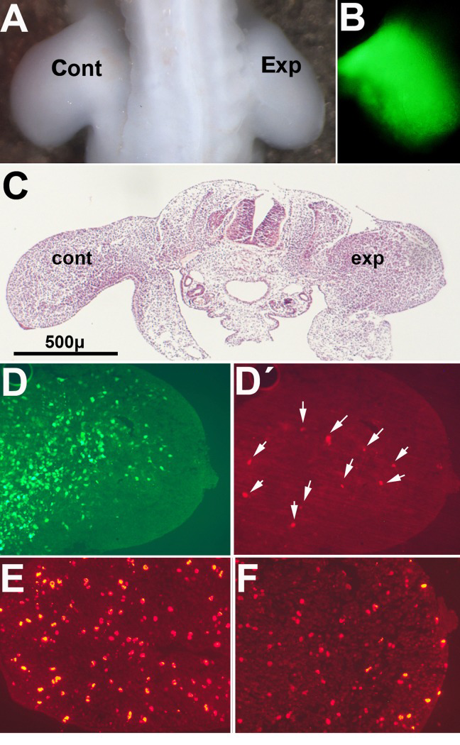
Figure 4.Pictures illustrate chick embryos 48 hr after electroporation of GFP /Btg2 constructs in the right wing bud at 2 id(A) Dorsal view of a chick embryo showing the reduced size of the experimental limb (Exp) compared with the contralateral control (Cont). (B) Mesodermal expression of GFP in the limb 48 hr after electroporation. (C) HE stained transverse section of an experimental embryo to show the different size of the experimental limb (exp) compared with the contralateral limb (cont). (D) and (D′), show the expression of GFP (D) and the presence of TUNEL-positive apoptotic cells (arrows in (D′) in a correlative section of the experimental limb in (C). E-F, are correlative sections of the embryo in (C) after immunolabeling with anti-p-H3. Note the reduced number of positive cells in the experimental limb (F) compared with the control limb (E).