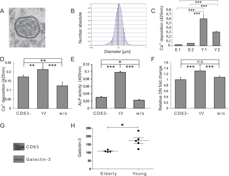
Figure 1.Vesicular impact on osteogenic differentiation capacity of ASCs(A) Electron microscopy picture of extracellular vesicles (EVs) isolated by differential centrifugation from human plasma. (B) Size distribution of plasma derived EVs analysed by nano tracking. (C) Mineralization of ASCs exposed to extracellular vesicles derived from donors older than 55 years (E1, E2) or from donors younger than 25 years (Y1, Y2) was evaluated by Alizarin Red staining. The released dye was quantified by microplate reader at 425nm. Mineralization was significantly increased in ASCs exposed to extracellular vesicles of young donors compared to ASCs exposed to vesicles of elderly donors. (D-F) Effects of CD63 positive plasma derived extracellular vesicles of young donors on osteogenesis of ASCs. (D) Mineralization of ASCs exposed to the CD63− fraction, the total EV fraction (tEV) or of unexposed ASCs was evaluated by Alizarin Red staining. The released dye was quantified by microplate reader at 425nm. Osteogenic differentiation was decreased when cells were exposed to the EV fraction depleted of CD63 positive vesicles. (E) Alkaline Phosphatase (ALP) activity was quantified by microplate reader at 405nm. Activity was significantly decreased when cells were exposed to the EV fraction depleted of CD63 positive vesicles. (F) Relative fold change of Osteonectin (ON) mRNA levels of ASCs was evaluated by qPCR and normalized to GAPDH. ON mRNA levels were significantly decreased when cells were exposed to the EV fraction depleted of CD63 positive vesicles. (C-F) ns: not significant, *: p<0.05, **: p<0.01, ***: p<0.001 in comparison to indicated group. Data are presented as mean values ± SD and were statistically analysed using 1-way ANOVA followed by a Bonferroni multiple comparison test, n=4. (G-H) Plasma derived vesicular Galectin-3 protein levels. (G) Detection of Galectin-3 and CD63 protein by Western blot in anti-CD63 immunopurified plasma derived extracellular vesicles. (H) Galectin-3 protein levels in extracellular vesicles of donors younger than 25 (Young) or older than 55 years (Elderly) were analysed by ELISA. Vesicular Galectin-3 protein levels significantly decrease with age. Grubbs' analysis identified an outlier in the elderly population (highlighted in red) who was excluded from subsequent statistical analysis.