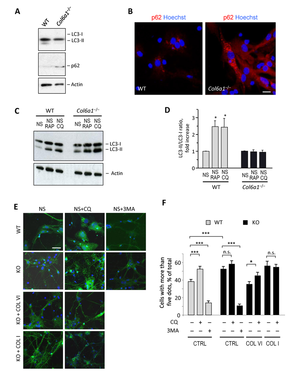
Figure 3.Col6a1−/− neural cell cultures display altered autophagic flux(A) Western blot analysis of LC3 and p62 in protein lysates from wild-type and Col6a1−/− primary neural cell cultures. Actin was used as a loading control. (B) Immunofluorescence for p62 (red) in wild-type and Col6a1−/− primary neural cultures, showing accumulation of p62 aggregates in Col6a1−/− cells. Nuclei were stained with Hoechst (blue). Scale bar, 100 μm. (C) Western blot analysis for LC3 in protein lysates from wild-type and Col6a1−/− primary neural cultures maintained for 4.5 hr in serum-free medium (NS), serum-free medium with 100 nM rapamycin (NS RAP), or serum-free medium with 50 μM chloroquine (NS CQ). Actin was used as a loading control. (D) Densitometric quantification of the LC3-II/LC3-I ratio, as determined by western blot analysis of three independent primary neural cultures from wild-type and Col6a1−/− mice. The LC3-II/LC3-I ratio is expressed as fold-change relative to the serum-free condition (*, P<0.05; n = 3). (E) Fluorescence microscopy analysis of GFP-LC3 distribution in neural cell cultures derived from GFP-LC3::Col6a1+/+ (WT) and GFP-LC3::Col6a1−/− (KO) mice and maintained for 4.5 hr in serum-free medium in the absence (NS) or in the presence of 50 μM chloroquine (NS+CQ) or of 10 mM 3-methyladenine (NS+3MA). Where indicated, before the experiment cells were seeded onto purified collagen VI (COL VI) or collagen I (COL I) as substrates. Nuclei were stained with Hoechst (blue). Scale bar, 50 μm. (F) Quantification of GFP-LC3 puncta in neural cell cultures derived from GFP-LC3::Col6a1+/+ (WT) and GFP-LC3::Col6a1−/− (KO) mice and maintained in the different conditions described above. The histogram shows the percentage of cells with more than five fluorescent puncta for each condition (***, P<0.001; *, P<0.05; n.s., not significant; n = 3). 3MA, 3-methyl-adenine; COL I, adhesion onto collagen I; COL VI, adhesion onto collagen VI; CQ, chloroquine; CTRL, adhesion onto poly-lysine; WT, wild-type.