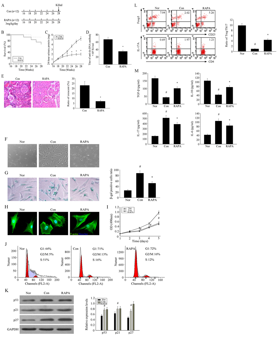
Figure 1.RAPA improves lupus nephritis by influencing the cellular senescence of BM-MSCs from MRL/lpr mice(A) The treatment group had intragastric administration of RAPA 3mg/kg/day between 16 and 28 weeks of ages. Survival curves observed that The survival rate of the RAPA-treated MRL/lpr mice was higher than that of control group. (B-E) We recorded the survival rate and the weight of mice. 24-hours urinary protein was measured by coomassie brilliant blue method. Mice were killed and were taken peripheral blood in orbit. Elisa showed that anti-ds-DNA antibody titer in serum in RAPA -treated group is lower than control group. HE-staining showed that renal pathological changes of control group is significant,including glomerular sclerosis, mesangial cell proliferation,matrix widened, and formation of crescent, a number of lymphocytes infiltrating the interstitium (HE×300). However, histopathological changes of RAPA -treated group were remarkable alleviated. (F) Cellular morphology observed that the RAPA-treated MSCs from MRL/lpr mice were less hypertrophic than untreated group (magnification; ×200). (G) MSCs were fixed and stained for β-gal. The number of SA-β-gal- positive cells among treated groups decreased in comparison with untreated group. (H) Immunofluorescence showed that the abnormal F-actin distribution in MSCs from MRL/lpr mice was reversed by RAPA treatment. (I) Cell Counting Kit (CCK)-8 method was used to detect the cell proliferation rate. (J) Flow cytometry was used to detect the distribution of cell cycle. (K) Western Blotting was used to detect the changes of cell cycle-related proteins. However, no remarkably differences were found between treated and untreated MSCs. (L) P4 MSCs transwell cultured with CD4+ T cells for 72 h. The count of Treg cells decreased and Th17 cells increased in MSCs from MRL/ lpr mice compared to the normal group by flow cytometry analysis. MSCs from RAPA-treated group could reverse the abnormal changes. The statistical results revealed that the treatment of RAPA could up-regulated the ratio of Treg/Th17 from MRL/lpr mice MSCs. (M) The supernatants of MSCs were collected. RAPA-treated group induced the secretion of IL-10 and TGF-β but reduced IL-17 and IL-6 by ELISA. All data were expressed as the mean±SEM (n = 3, *P<0.05 compared with normal group, #P<0.05 compared with the untreated group).