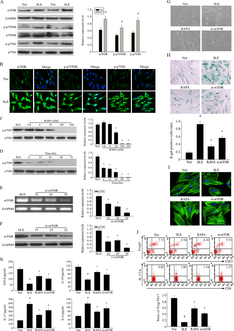
Figure 3.Over-activation of mTOR pathway was involved in the senescence of MSCs from SLE patients(A) The over-expression of p-mTOR, p-S6K and p-S6 in MSCs from SLE compared with normal group were determined by western blot analysis. GAPDH was used as the internal control. (B) P4 MSCs from SLE patients and normal group were cultured in 24-well plates. Immunofluorescence staining of p-mTOR, p-S6K and p-S6 verified their over-activation in SLE MSCs. Counterstaining with DAPI displays the localization of the nucleus (Scale bar = 50 μm). (C-D) P4 MSCs from SLE patients cultured at the different concentration of RAPA for 72 h. RAPA achieved maximal effects at about 500 nM by assaying the inhibition of p-70S6. (E-F) BMMSCs were depleted of mTOR by RNAi. The third one was chosen as the best siRNA by western blotting. (G) P4 MSCs from SLE patients were treated with 500 nM RAPA and si-mTOR or not for 72 h. Cellular morphology showed that the RAPA and si-mTOR treated SLE MSCs were less hypertrophic than untreated group (magnification; ×200). (H) MSCs were fixed and stained for β-gal. The number of SA-β-gal- positive cells obviously decreased among treated SLE MSCs in comparison with untreated group. (I) Immunofluorescence showed that RAPA and si-mTOR reversed abnormal F-actin distribution in MSCs from SLE patients. (J) P4 SLE MSCs were treated with 500 nM RAPA and the third si-mTOR or not, then transwell cultured with CD4+T cells for 72 h. The count of Treg cells decreased and Th17 cells increased in SLE MSCs compared to the normal group by flow cytometry analysis. But si-mTOR and RAPA-treated MSCs could reverse the abnormal changes. The statistical results revealed that RAPA could up-regulated the ratio of Treg/Th17 from SLE MSCs, which was down-regulated compared with normal group. (K) The supernatants of MSCs were collected. si-mTOR RAPA-treated SLE MSCs induced the secretion of IL-10 and TGF-β but reduced IL-17 and IL-6 by ELISA. All data were expressed as the mean±SEM (n = 3, *P<0.05 compared with normal group, #P<0.05 compared with the untreated group).