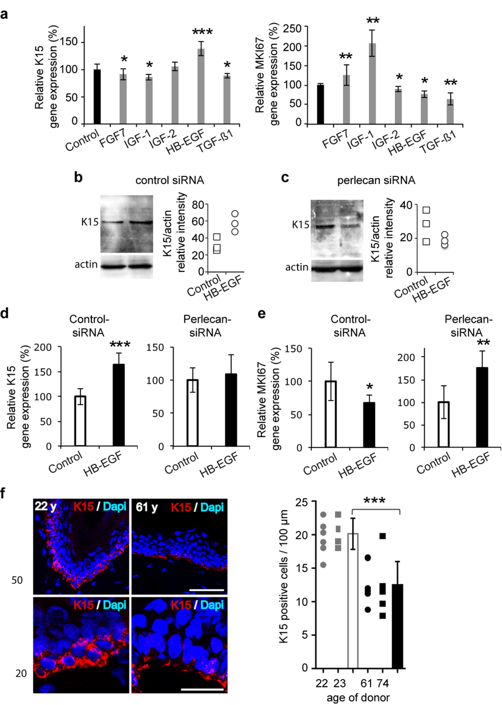
Figure 4.Perlecan functions in K15 expression regulation in aging skin(a) Real time PCR analysis of KRT15 and MKI67 in the 52‐year old keratinocyte strain after treatment with growth factors (FGF7, 15 ng/ml; IGF‐1, 50 ng/ml; IGF‐2, 80 ng/ml; HB‐EGF, 100 ng/ml; TGF‐Δ1, 1 ng/ml). (b,c) Western blot of K15 expression in control (b) and perlecan knock‐down (c) keratinocytes after HB‐EGF treatment (100 ng/ml). Quantifications of K15 expression in control‐siRNA and perlecan‐siRNA transfected cells are relative to actin expression. Experiment was done in triplicate. (d) Real time PCR analysis of KRT15 and (e) MKI67 in control and perlecan knock‐down keratinocytes after HB‐EGF treatment (100 ng/ml). (f) K15 (red) immunostaining and DAPI (blue) staining in 2 young and 2 aged skin biopsy sections. Red and blue cells were counted along the BM of 6 independent sections. Scale bars = 50 μm (top) and 20 μm (bottom). (a, d, e). Data are representative of at least 12 independent experiments with keratinocytes from the 52 years old donor. Mean ± SD; *p<0.025,**p<0.005, ***p<0.0005 vs. control, Student's t‐test.