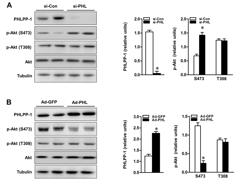
Figure 3.PHLPP-1 expression level affected Akt phosphorylation at Ser473(A) Cardiomyocytes were transfected with PHLPP-1-specific (si-PHL) and scrambled negative control (si-Con) siRNA for 48 hours. Representative immunoblots demonstrating the two sites of Akt phosphorylation with PHLPP-1 knockdown (left panel). The bar graphs (right panel) show the relative levels of PHLPP-1 and Akt phosphorylation (S473 & T308, normalized by total Akt bands), respectively. (B) Cardiomyocytes were infected with the recombinant human-phlpp-1 over-expression plasmid (Ad-PHL) and negative control (Ad-GFP) adenovirus for 72 hours. Representative immunoblots demonstrating the two sites of Akt phosphorylation with PHLPP-1 upregulation (left panel). The bar graphs (right panel) show the relative levels of PHLPP-1 and Akt phosphorylation (S473 & T308), respectively (*P < 0.05 vs. respective negative control. Values are means ± S.E., n = 5 per group).