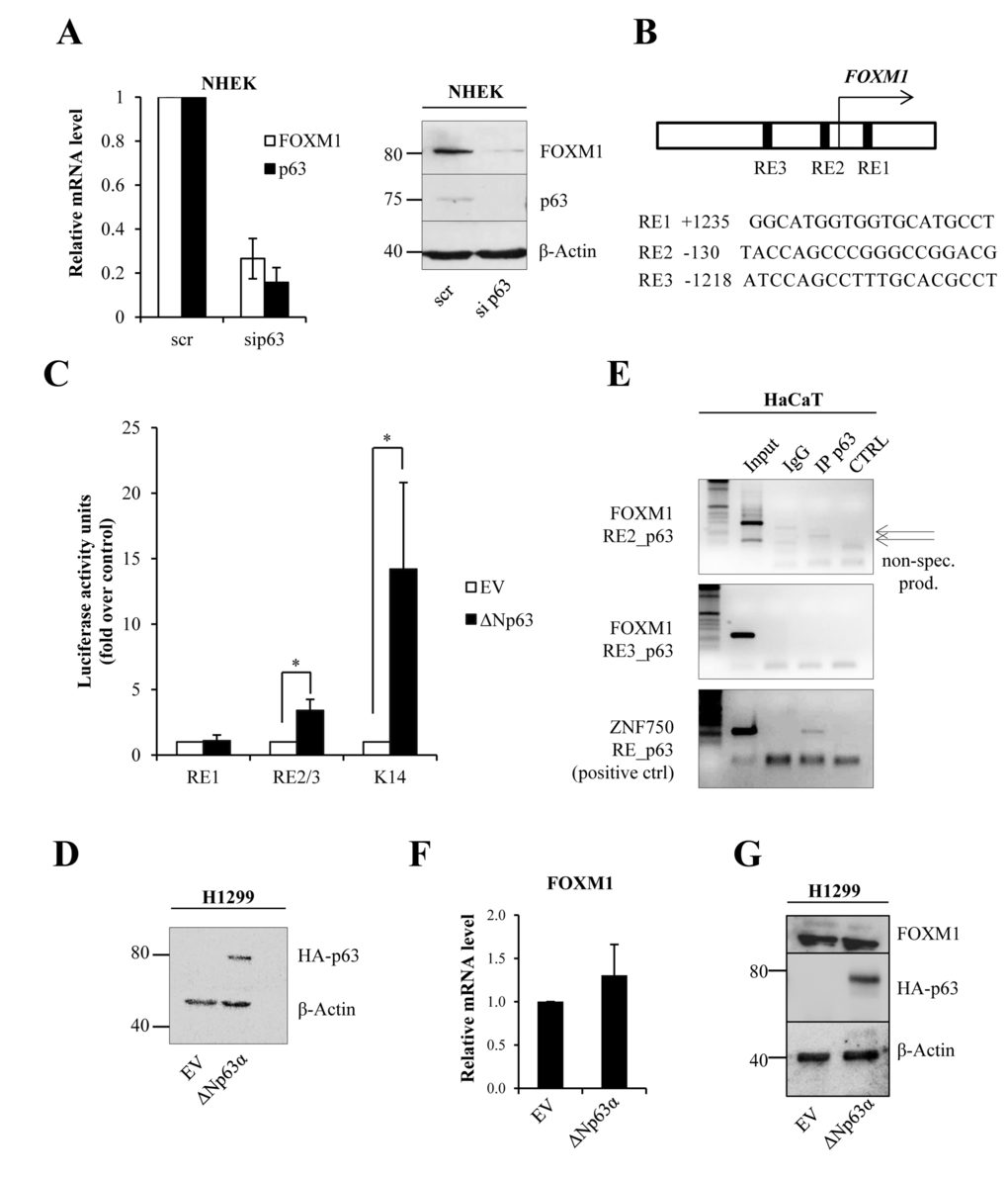
Figure 1.ΔNp63 indirectly regulates FOXM1 expression(A)NHEKs were silenced for p63, and the relative mRNA (48 h after transfection) and protein (96 h after transfection) levels of FOXM1 and p63 were determined. Values reported are the average ± SD of three independent experiments. (B) Scheme of the FOXM1 promoter showing the identified putative p63 response elements (REs). (C) Luciferase activity assay in H1299 cells transfected with pGL3 vectors containing putative response elements from the FOXM1 promoter. Values reported are the average ± SD of three independent experiments. *p-value <0.05 by Student's t-test. (D)Western blot analysis of the lysates that were used for luciferase assays. (E)Chromatin immunoprecipitation was performed in HaCaT cells with anti-p63 antibody (IP p63) or negative control immunoglobulin G (IgG). PCR was carried out with specific primers for putative p63 response elements in the FOXM1 promoter (RE2 and RE3). The ZNF750 promoter was used as a positive control for immunoprecipitation. The arrows indicate non-specific products. (F)H1299 cells were transfected with pcDNA vector expressing ΔNp63α–HA. After 24 h, FOXM1 expression levels were analyzed by qPCR. (G)Western blot showing ΔNp63α–HA expression and FOXM1 level.