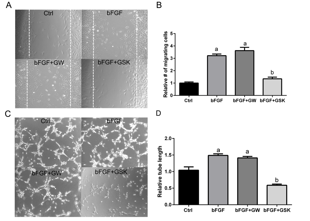
Figure 6.Antagonism of PPARβ/δ blocks endothelial cell migration and tube formation. (A) The effect of ligand activation or antagonism of PPARβ/δ on migration of RF/6A cells was analyzed in a bFGF induced wound-healing assay (n = 3, representative images at t = 36 hours are shown); dotted lines demarcate the boarders of the scrape wound. Ctrl: media only, bFGF: basic fibroblast growth factor (10 μg/ml), bFGF+GW: bFGF plus GW0742 (10μM), bFGF+GSK0660: bFGF plus GSK0660 (10 μM). (B) The cells migrating into the wound were counted using ImageJ (mean and S.E.M.; n = 3; a, p < 0.01 relative to Ctrl; b, p < 0.01 relative to bFGF, one way ANOVA, Tukey’s multiple comparisons test). (C) The effect of ligand activation or antagonism of PPARβ/δ on bFGF-induced tube formation in RF/6A cells was analyzed by an angiogenesis assay in Geltrex™ (n = 3; representative images at t = 3 hours are shown). Ctrl: media only, bFGF: basic fibroblast growth factor (10 μg/ml), bFGF+GW: bFGF plus GW0742 (10 μM), bFGF+GSK0660: bFGF plus GSK0660 (10 μM). Suramin, an inhibitor of tube formation, was used as a negative control (data not shown). (D) Quantification of tube length in Geltrex™ using ImageJ (mean and S.E.M.; n = 3; a, p < 0.01 relative to Ctrl; b, p < 0.01 relative to bFGF, one way ANOVA, Tukey’s multiple comparisons test).