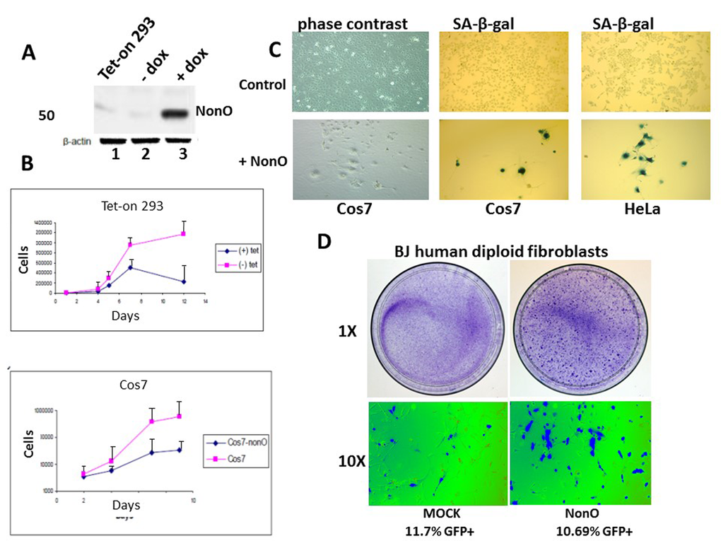
Figure 1.Overexpression of NonO promotes senescence of transformed and primary cells. (A) Cell lysates from Tet-on 293 (lane 1), a clone stably transfected with NonO in the presence (lane 3) and absence (lane 2) of tetracycline, were confirmed for OE by SDS-PAGE/anti-NonO western blotting. Error bars represent average of 3 independent measurements. (B) Growth rate retardation following NonO overexpression. An inducible clone of Tet-on 293 (upper panel) or Cos7 (lower panel) was plated on day 0 at 10,000 cells/well in the presence (+) and absence (-) of tetracycline and harvested for cell counting as plotted as a function of time on a log scale. Error bars represent average of 3 independent measurements. (C) NonO overexpression promotes senescence of 293T and Cos7 cells, as measured by SA-β-gal staining. Empty vector (mock, upper panels); NonO overexpression (OE, bottom panels). Photographs are at the same magnification. (D) Overexpression of GFP-NonO promotes senescence of human diploid BJ fibroblasts. SA-β-gal staining indicated in blue of mock (left) and GFP-NonO (right) at day 12 following stable transfection/G418 selection. Magnification of 1 and 10X is indicated to the left of respective panels. Equivalent transfection efficiencies, indicated at bottom by %GFP+, were confirmed by flow cytometry analysis (not shown).