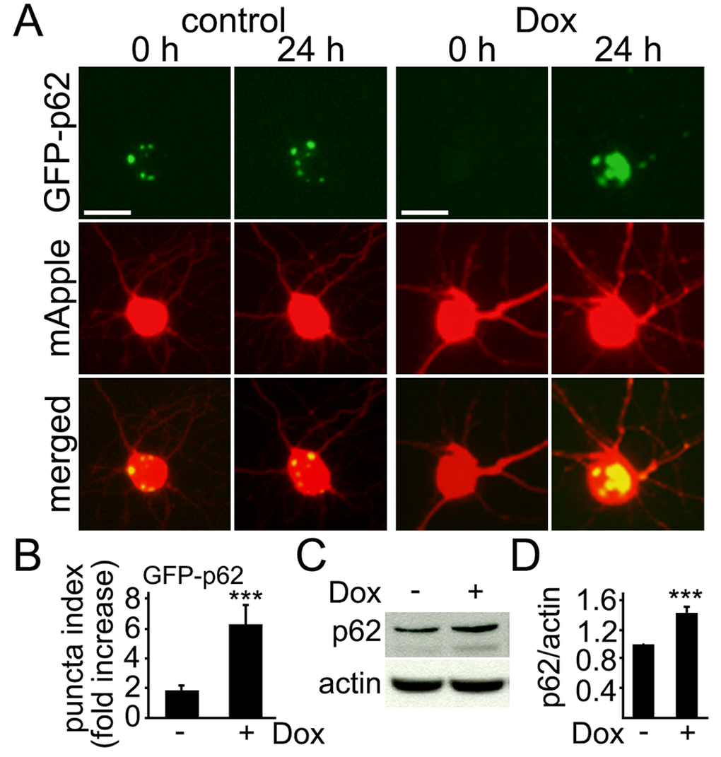
Figure 2.Levels of p62 increase in cultured cortical neurons treated with doxorubicin. (A) Autophagy was impaired by doxorubicin. Cortical neurons were transfected with mApple (a morphology and viability marker) and GFP-p62. The first neuronal cohort was treated with a vehicle, and the second cohort was treated with 50 nM doxorubicin (overnight). Neurons were imaged before and after the treatments. Small aggresomes are sometimes formed in neurons. Note large inclusion bodies formed by GFP-p62 in doxorubicin-treated neurons. Bar, 10 μm. (B) Quantification of fluorescent images from (A). The fold-increase of the puncta index in neurons, which express GFP-p62, treated with a vehicle or with 50 nM doxorubicin (overnight). *** p<0.0001 (t-test). Two hundred neurons were analyzed from two independent experiments. (C) Endogenous p62 accumulated in cultured cortical neurons treated with 50 nM doxorubicin (overnight). Actin was used as a loading control. (D) Quantification of western blots from (C). The levels of p62 were normalized to actin. *** p<0.0001 (t-test). Results were pooled from four independent experiments.