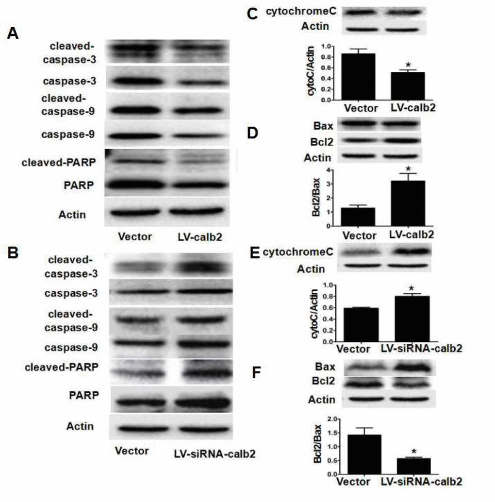
Figure 4.Calretinin inhibits the apoptosis of Leydig cells partially via mitochondrial-related apoptotic pathways. After MLTC-1 cells and R2C cells were transfected with LV-calb2, LV-siRNA-calb2 or vector alone, the expression of factors in the mitochondrial-related apoptotic pathways was analyzed by Western blotting. (A) In MLTC-1 cells with up-regulated calretinin expression, thecaspase-3/9, cleaved caspase-3/9, PARP, and cleaved-PARP expression levels were decreased. (B) In R2C cells with down-regulated calretinin expression, the caspase-3/9, cleaved caspase-3/9, PARP, and cleaved-PARP expression levels were increased. (C) Cyto C expression was significantly lower in MLTC-1 cells with up-regulated calretinin. (D) The Bcl2/Bax ratio was significantly increased in MLTC-1 cells with up-regulated calretinin expression. (E) In R2C cells with the down-regulated calretinin, cyto C expression was significantly increased. (F) In R2C cells with down-regulated calretinin, the Bcl2/Bax ratio was significantly lower. The vector was used as the negative control(s). *: p<0.05.