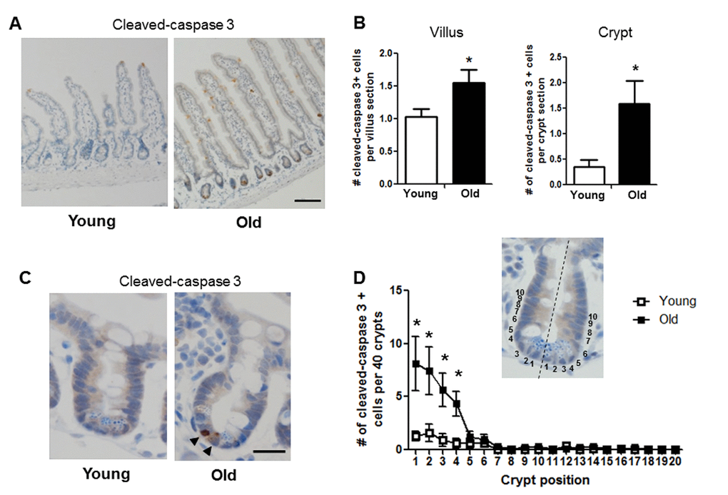
Figure 6.Increased proportion of small intestinal epithelial cell apoptosis with age. (A) Representative image of crypt and villus sections stained for cleaved caspase-3. Magnification: 10x, Scale bar: 100µm . (B) Quantification of the number of cleaved caspase-3 positive cells per villus section (n=7 animals per group) and per crypt section (n=10 young and 9 old animals). *p<0.05 Young vs. Old, unpaired t test. (C) Representative image of crypt sections stained for cleaved caspase-3. Magnification: 40x, Scale bar: 20µm. (D) Quantification of the location of cleaved caspase-3 positive cells per 40 crypts per animal by position. Inlay shows method of identifying cell position within crypt. n=10 young and 9 old animals, *p<0.05 Young vs. Old, unpaired t test.