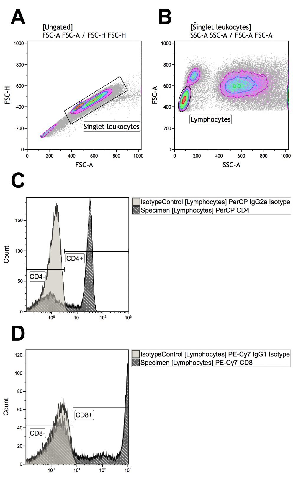
Figure 1.Gating strategies for identifying T-cells that are CD4+ (defined as CD4+CD8-) or CD8+ (defined as CD4-CD8+). (A) Singlet leukocytes were identified using forward scatter height vs. area scatter on a combined contour and density plot. (B) Lymphocytes were identified using forward scatter area vs. side scatter area on a combined contour and density plot. (C) CD4+ and CD4- lymphocytes were identified with the help of negative isotype controls (set at 1% to distinguish fluorescence signal from non-specific background signals). (D) CD4+ and CD4- lymphocytes were identified with the help of negative isotype controls (set at 1% to distinguish fluorescence signal from non-specific background signals). Using logical Boolean sequences, we distinguished two T-cell populations for further analyses: CD4+ T-cells that were CD4+CD8- and CD8+ T-cells that were CD4-CD8+.