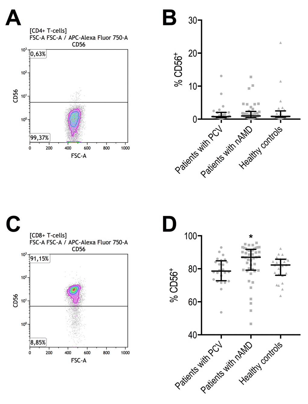
Figure 4.Proportion of CD56+ CD4+ and CD8+ T-cells in patients with polypoidal choroidal vasculopathy (PCV), patients with neovascular age-related macular degeneration (nAMD), and healthy controls. (A) CD56+ CD4+ T-cells were identified with the help of negative isotype controls (set at 1% to distinguish fluorescence signal from non-specific background signals). (B) CD56+ in CD4+ T-cells did not differ between the groups. Horizontal line with whiskers indicate median and interquartile range. (C) CD56+ CD8+ T-cells were identified with the help of negative isotype controls (set at 1% to distinguish fluorescence signal from non-specific background signals). (D) CD56+ in CD8+ T-cells differed significantly and was significantly higher in patients with nAMD (signified with *, P = 0.0016, Kruskal-Wallis test).