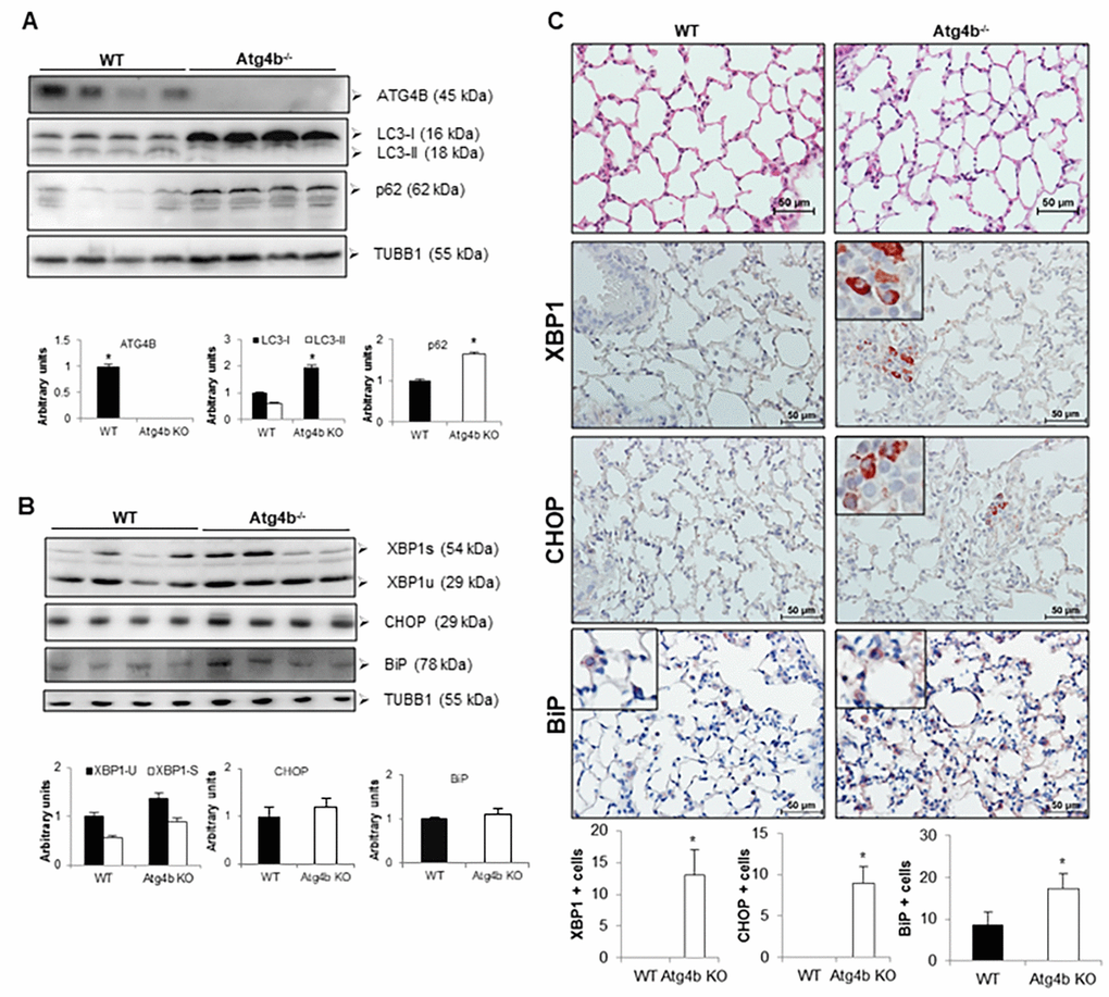
Figure 3.Loss of autophagy function by Atg4b deficiency resulted in mild ER stress induction. (A) Representative immunoblots of ATG4B, LC3-I/ II, and p62 in lung tissue from WT and Atg4b null mice in basal conditions. (B) Representative immunoblots of ER stress biomarkers in lung tissue from WT and Atg4b null mice in basal conditions. β-tubulin was used as loading control. Densitometry analysis (bottom panels). (C) Representative photomicrographs of immunohistochemical staining performed with specific primary antibodies against XBP1, CHOP and BiP in lung tissues sections from WT and Atg4b in unchallenged/basal conditions. Positive signal is observed in red. All sections were counterstained with hematoxylin. All insets show 2X larger magnification. Total number of positive stained cells per high power field (40X) in lung tissue sections by quantitative image analysis. Results are shown as mean ± SD. Statistical significance was determined by Student´s t-test (*p < 0.05).