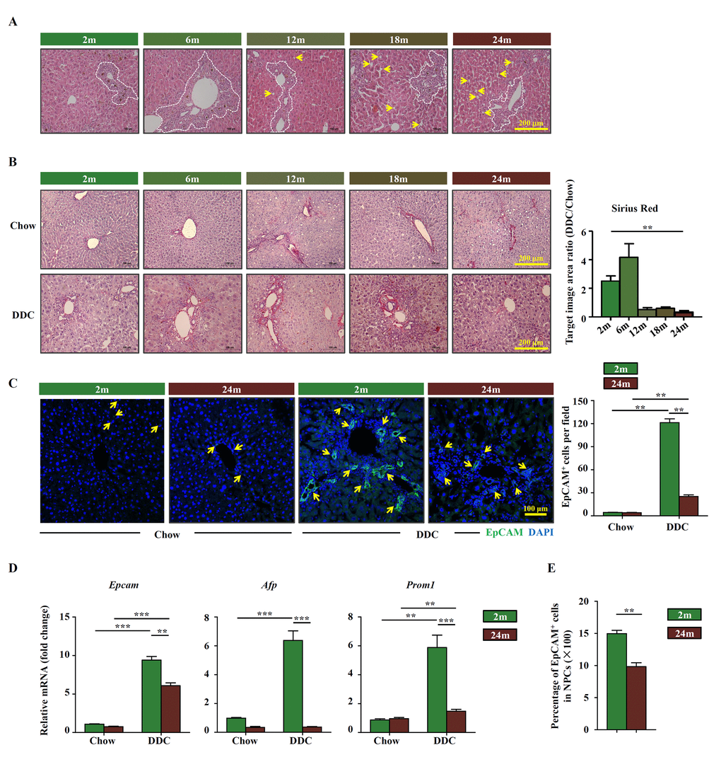
Figure 1.OC activation is impaired in aged mice. (A) H&E staining of DDC-fed mouse liver with the age of 2m, 6m, 12m, 18m and 24m (Scale bar=200 μm). The ductular reaction was shown in the white dashed lines outlined regions. Arrows indicated the hepatocyte steatosis. (B) Sirius red staining of the chow (Chow) and DDC groups (DDC) with the age of 2m, 6m, 12m, 18m and 24m (Scale bar=200 μm). The ratio of Sirius red staining area of DDC and chow groups was quantified (n=6, ** p < 0.01). Young (2m) and aged (24m) mice were fed with normal/DDC diet for 3 weeks. (C) Immunofluorescence staining for EpCAM+ cells (green) in DDC-fed (DDC) versus chow controls (Chow). Quantification of EpCAM+ cells was shown (n=6, ** p < 0.01). (D) Quantitative Real-time PCR analysis of Epcam, Afp and Prom1 in young and aged DDC-fed mice liver (n=9, ** p < 0.01, *** p < 0.001). (E) EpCAM+ cells and NPCs were isolated from whole liver of DDC-fed young and aged mice, the ratio of EpCAM+ cells in NPCs was quantified (n=6, ** p < 0.01).