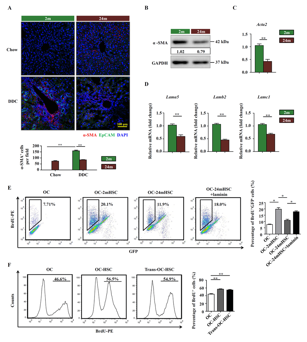
Figure 4.HSCs participate in the remodeling of OC niche by producing laminin. (A) Immunofluorescence staining for α-SMA+ (red) and EpCAM+ (green) cells in DDC-fed (DDC) versus chow controls (Chow) of young (2m) and aged (24m) mice. Quantification of α-SMA+ cells was shown (n=6, ** p < 0.01). (B) Western blot analysis of α-SMA in HSCs freshly isolated from young (2m) and aged (24m) mice with DDC diet. (C) Quantitative Real-time PCR analysis of Acta2 in HSCs freshly isolated from young (2m) and aged (24m) mice with DDC diet (n=6, ** p < 0.01). (D) Quantitative Real-time PCR analysis of different laminin isotypes in HSCs freshly isolated from young and aged mice with DDC diet (n=6, ** p < 0.01). (E) BrdU analysis of OCs co-cultured with primarily isolated HSCs from young (OC-2mHSC) and aged (OC-24mHSC) GFP transgenic mice while in one group laminin was added (OC-24mHSC+laminin) (n=4, * p < 0.05). (F) BrdU analysis of OCs co-cultured with HSCs in a cell-cell contact manner (OC-HSC) or with a transwell system (trans-OC-HSC) (n=5, ** p < 0.01).