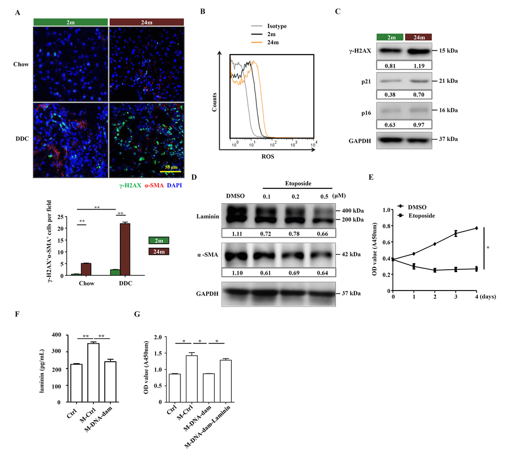
Figure 5.Aged HSCs provide less support to OCs. (A) Immunofluorescence staining for γ-H2AX+ (green) and α-SMA+ (red) cells in DDC-fed (DDC) versus chow (Chow) of young (2m) and aged (24m) mice. Quantification of γ-H2AX+α-SMA+ cells was shown (n=6, ** p < 0.01). (B) ROS levels were analyzed in HSCs freshly isolated from young (2m) and aged (24m) mice with DDC diet. (C) Western blot analysis of indicated proteins in HSCs freshly isolated from young (2m) and aged (24m) mice with DDC diet. (D) Western blot analysis of indicated proteins in JS1 cells pretreated with different doses of etoposide for 24 hours. (E) CCK-8 test showed the proliferation of JS1 cells treated with etoposide (0.5 μM; n=5, * p < 0.05). (F) The level of laminin in normal culture condition (Ctrl), conditional medium of HSC supernatant (M-Ctrl), and conditional medium of etoposide pretreated HSC supernatant (M-DNA-dam) was measured by ELISA (n=5, ** p < 0.01). (G) CCK-8 test shows that proliferation of OCs in conditional medium with ordinary culture condition (Ctrl), HSC supernatant (M-Ctrl), or conditional medium of etoposide pretreated HSC supernatant (M-DNA-dam), or in the presence of laminin (M-DNA-dam-Laminin) (n=4, * p < 0.05).