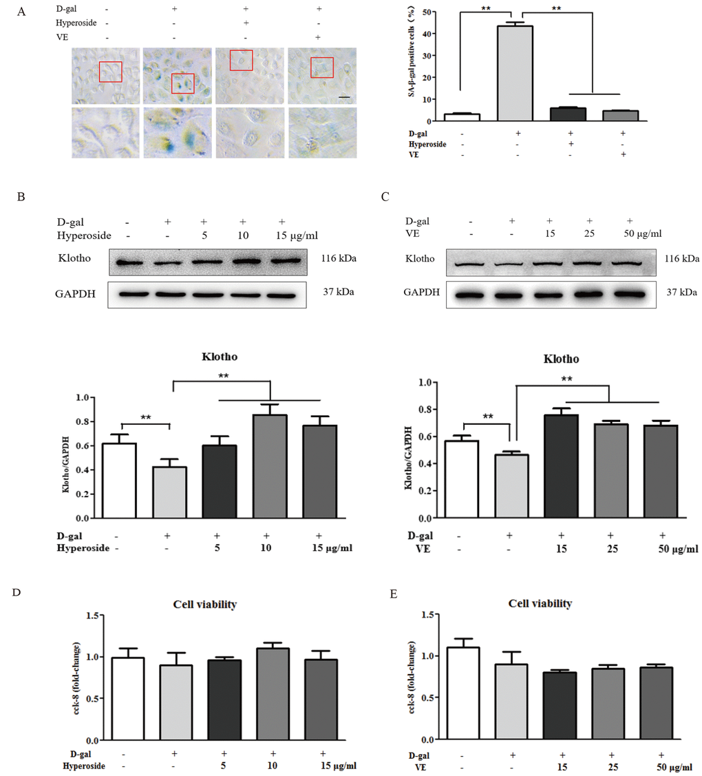
Figure 3.The effects of hyperoside and vitamin E on renal cellular aging and injury in vitro. (A) SA-β-gal staining in the NRK-52E cells exposed to D-gal at 100 mM, with the treatment of hyperoside at 10 μg/ml or VE at 25 μg/ml for 24 hours, and the percentage of SA-β-gal-positive cells. (B, C) The NRK-52E cells were exposed to D-gal at 100 mM, with the treatment of hyperoside at 0, 5, 10, and 15 μg/ml and VE at 0, 15, 25, and 50 μg/ml for 24 hours, and subjected to a WB analysis for klotho, respectively. (D, E) The cell viability in the NRK-52E cells exposed to D-gal at 100 mM, with the treatment of hyperoside at 0, 5, 10, and 15 μg/ml and VE at 0, 15, 25, and 50 μg/ml for 24 hours. The data are expressed as the mean ± SD, (n=3), **P < 0.01. Abbreviation: SA-β-gal, senescence-associated-β-galactosidase; D-gal, D-galactose; VE, vitamin E, WB, Western blot.