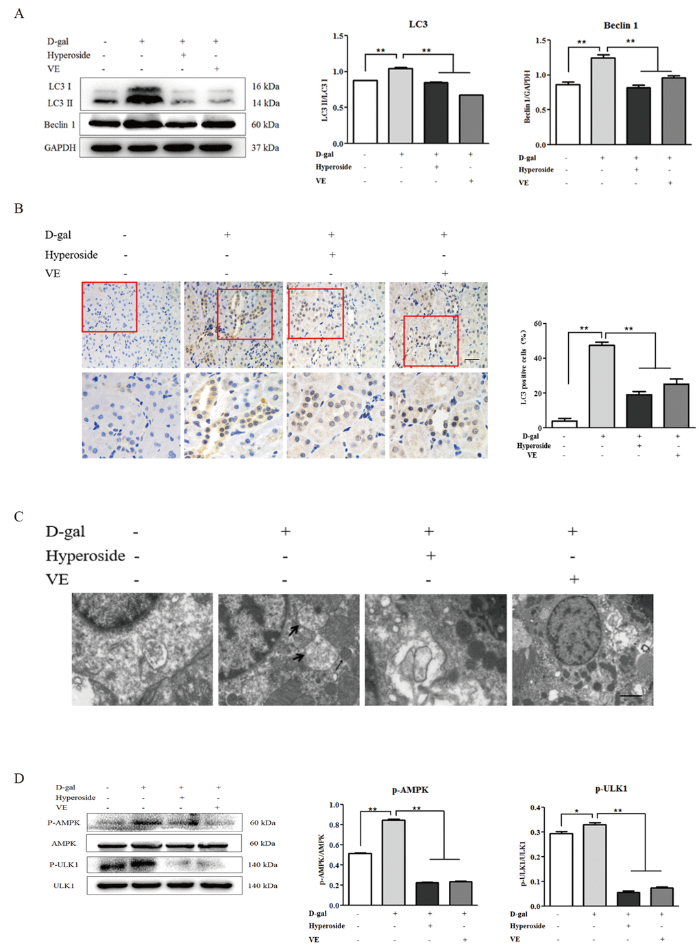
Figure 6.The effects of hyperoside and vitamin E on autophagic activity and the AMPK-ULK1 signaling pathway in vivo. (A) A WB analysis of LC3 I/II and Beclin1 in the kidneys from the rats in the control, the 8 week-D-gal, the D-gal + Hyperoside and the D-gal + VE groups. (B) Immunohistochemical staining of LC3 and the percentage of the positively stained areas of LC3 in the control, the 8 week-D-gal, and the D-gal + Hyperoside groups. Scale bar = 20 μm. (C) The morphological changes in the renal tubular cells of the rats in the control, the 8 weeks-D-gal, the D-gal + Hyperoside and the D-gal + VE groups by transmission electron microscopy. The black arrows show the autophagosomes with the characteristic morphology of a double membrane. (D) A WB analysis of p-AMPK, AMPK, p-ULK1 and ULK1 in the kidneys of the rats in the control, the 8 weeks-D-gal, the D-gal + Hyperoside and the D-gal + VE groups. The data are expressed as the mean ± SD, (n=3), *P < 0.05, **P < 0.01. Abbreviation: WB, Western blot; D-gal, D-galactose; VE, vitamin E; p-AMPK, phosphorylated AMPK; p-ULK1, phosphorylated ULK1.