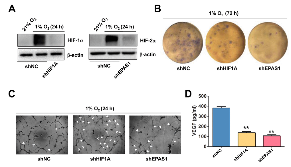
Figure 3.Knockdown of either HIF-1α or HIF-2α impairs hypoxia-induced angiogenesis and VEGF production in ECs. (A) HUVECs were transfected with pLKO.1 constructs encoding shRNA specifically targeting HIF-1α and HIF-2α, as well as scramble sequence as negative control (shNC). As the basal levels of HIF-1α or HIF-2α in HUVECs were relatively low, these transfected cells were exposed to 1% O2 (21% O2 as normoxia control) for 24 hrs, after which Western blot analysis was performed to confirm shRNA knockdown of HIF-1α and HIF-2α, respectively. (B, C) HUVECs expressing HIF-1α or HIF-2α shRNA were then exposed to 1% O2 for the indicated intervals, followed by colony formation assay (B, 72 hrs) and Matrigel-based tube formation assay (C, 24 hrs). Representative microscopic images for at least three independent experiments were shown. Arrowheads indicate unclosed loops of vascular structure. (D) In parallel, the VEGF level was measured by ELISA assay after cultured for 72 hrs under 1% O2. Values represent the means ± SD for at least three independent experiments performed in triplicate. **P < 0.01 for comparison with shNC control.