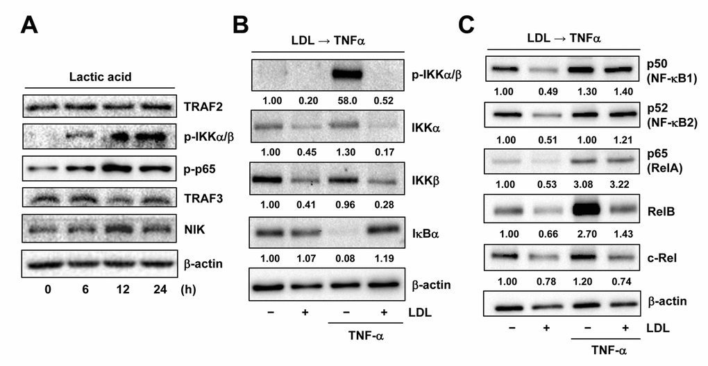
Figure 6.LDL inactivates TNFα-induced NF-κB activation via down-regulation of its key signaling components. (A) HUVECs were treated with 3 mM lactic acid as described in Figure 2D, after which Western blot analysis was performed to assess activation of the canonical (e.g., TRAF2 expression as well as IKKα/β and p65 phosphorylation) and non-canonical (e.g., expression of TRAF3 and NIK) NF-κB pathways. (B, C) HUVECs were pre-treated with LDL (100 μg/ml) for 48 hrs, followed by TNFα (50 ng/ml) for additional intervals as below, after which Western blot analysis was carried out to monitor phosphorylation of IKKα/β (Ser176/180, 5 min) as well as expression of multiple key components of both canonical and non-canonical NF-κB pathways, including IKKα, IKKβ, IκΒα (5 min), p50 (NF-κB1), p52 (NF-κB2), p65 (RelA), RelB, and c-Rel (4 hrs). All blots were densitometrically quantified, and values indicate fold increase after normalization to β-actin.