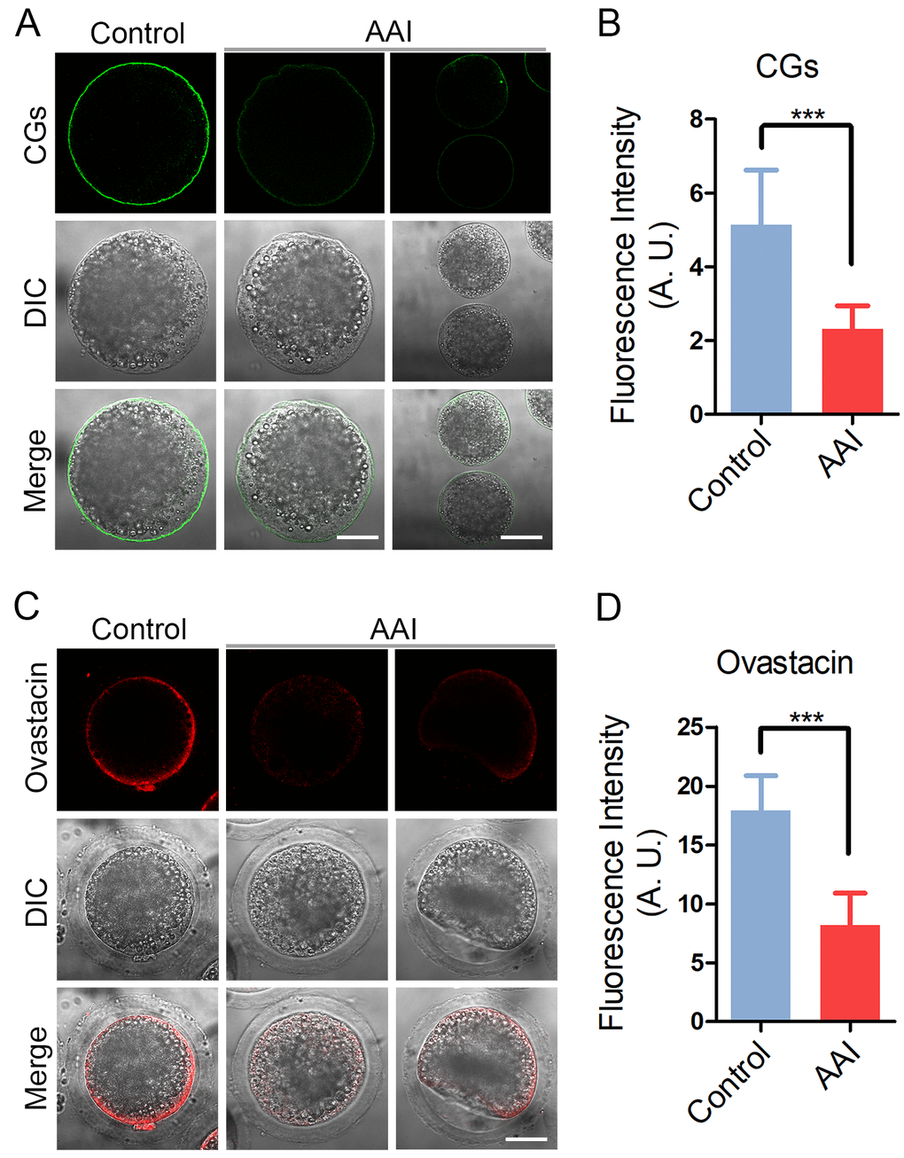
Figure 6.Effects of AAI exposure on the distribution of cortical granules and ovastacin in porcine oocytes. (A) Representative images of cortical granule localization in control and AAI-exposed oocytes. MII oocytes cultured for 44 h in vitro were stained with LCA-FITC to display the cortical granules. Scale bar, 30 and 60 μm. (B) The fluorescence intensity of cortical granules was measured around the signals on the plasma membrane in control and AAI-exposed oocytes. (C) Representative images of ovastacin localization in control and AAI-exposed oocytes. Ovastacin was immunostained with rabbit polyclonal anti-human ovastacin antibody and imaged by confocal microscope. Scale bar, 30 μm. (D) The fluorescence intensity of ovastacin was measured in control and AAI-exposed oocytes. Data in (B) and (D) were presented as mean percentage (mean ± SEM) of at least three independent experiments. ***P < 0.001.