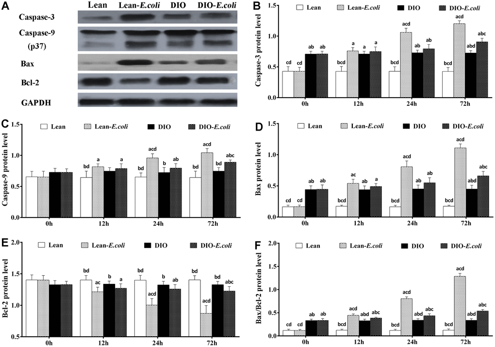
Figure 10.The changes of protein levels in the mice liver after E. coli infection. (A) The representative image of apoptotic regulatory proteins at 72h after receiving intranasal instillations. (B–F) Quantitative analysis of the relative protein expression (expressed as fold change relative to the lean group). Note: Letter a, b, c or d represent difference (p<0.05) between the group and the lean group, lean-E. coli group, DIO group, or DIO-E. coli group, respectively.