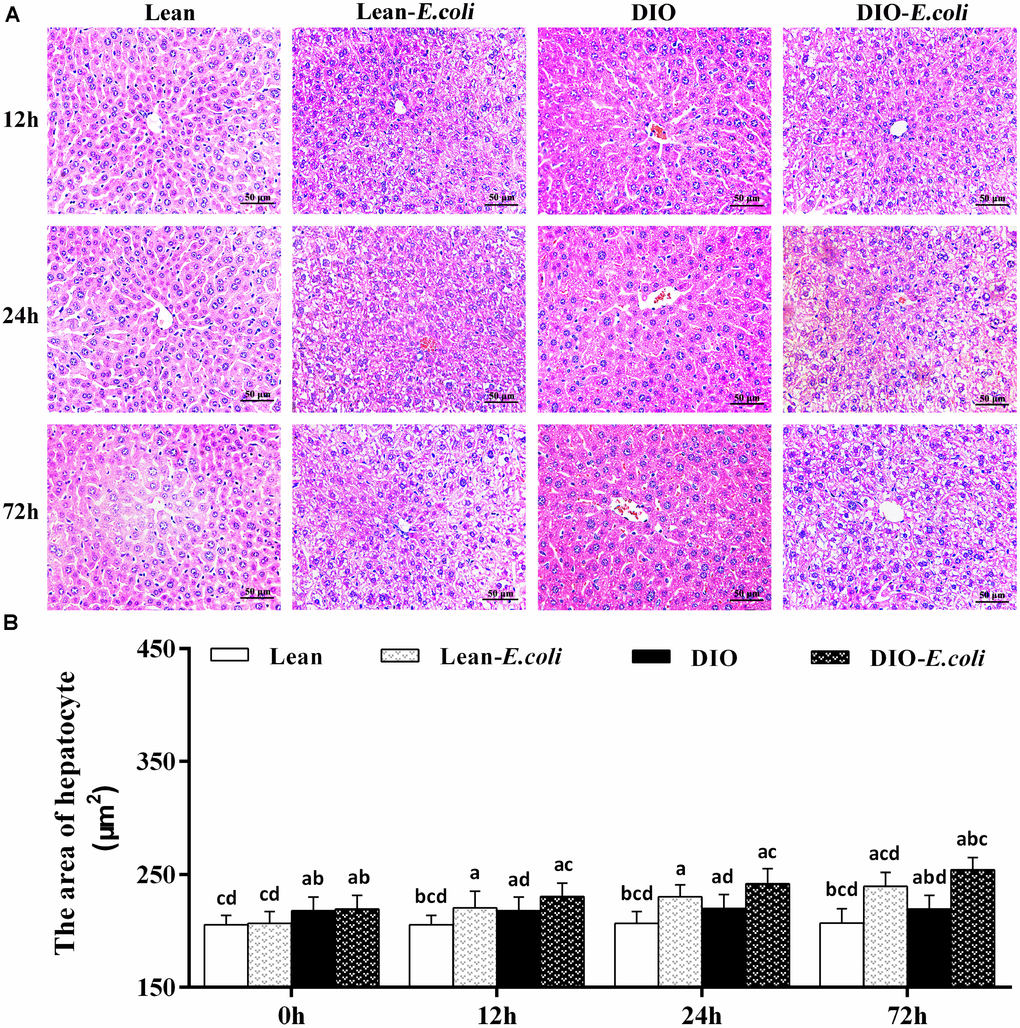
Figure 3.The histopathological changes of the liver after E. coli infection. (A) The representative histopathological images of livers (HE staining, bar=50 μm); (B) The area of hepatocyte. Note: Letter a, b, c or d represent difference (p<0.05) between the group and the lean group, lean-E. coli group, DIO group, or DIO-E. coli group, respectively.