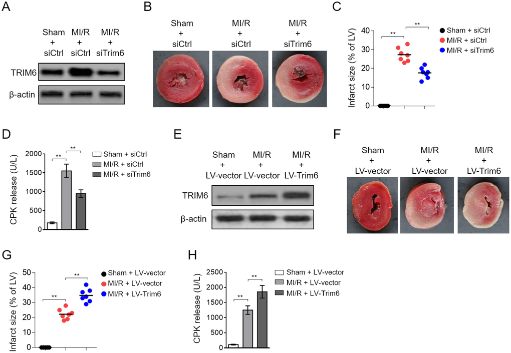
Figure 2.Cardiac TRIM6 promotes MI/R injury. (A–D) The mouse heart was pre-transfected in vivo with control siRNA (siCtrl) or siRNA targeting Trim6 (siTrim6) 48 hrs before surgery. Mice were then subjected to sham surgery or experimental MI/R. Each group contained 7 mice. At 24 hrs after reperfusion, the heart and serum samples were harvested for analyses. (A) The protein level of TRIM6 in the heart was determined by Western blotting analysis. β-Actin was used a loading control. (B) The mid-myocardial cross sections of hearts were stained with TTC to show infarct size (IS). The representative images are shown. (C) The infarct severity in each group shown as in (B) is expressed as % of LV (IS/LV). (D) The level of serum creatine phosphokinase (CPK) from each group was measured (U/L). (E–H) The mouse heart was pre-infected in vivo with lentivirus expressing vector control (LV-vector) or Trim6 (LV-Trim6) 48 hrs before surgery. Mice were then subjected to sham surgery or experimental MI/R. Each group contained 7 mice. At 24 hrs after reperfusion, the heart and serum samples were harvested for analyses. (E) The protein level of TRIM6 in the heart was determined by Western blotting analysis. β-Actin was used a loading control. (F) The mid-myocardial cross sections of heart were stained with TTC. The representative images are shown. (G) The quantification of IS in each group (% of LV). (H) The level of serum creatine phosphokinase (CPK) from each group was measured (U/L). All data are expressed as mean ± SD (n = 7). **, P < 0.01.