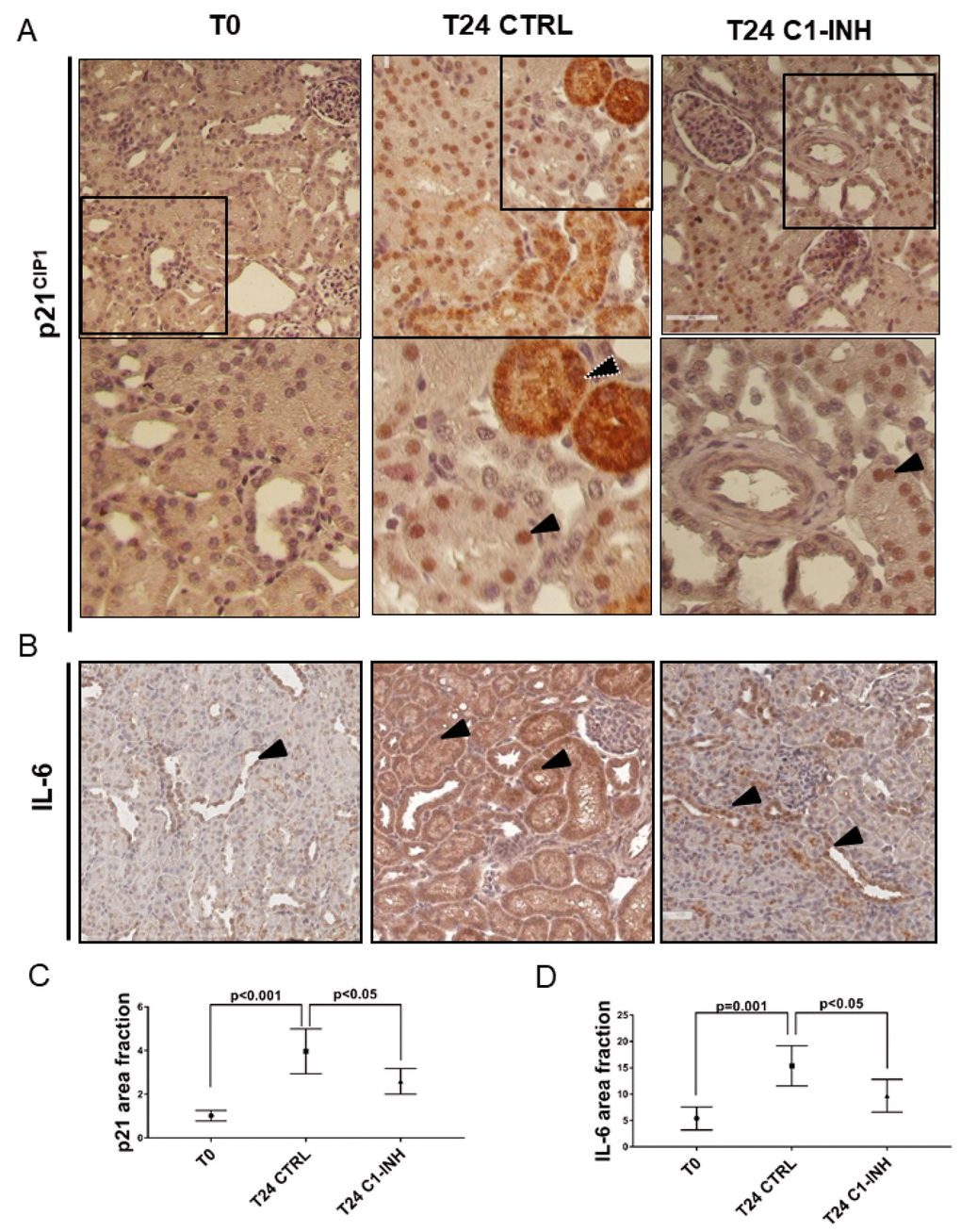
Figure 8.p21 and IL-6 are induced in tubular cells after I/R and modulated by C1-INH treatment. (A) Representative micrographs indicating p21/CIP1 protein expression in different groups of swine after I/R injury, as indicated. Boxed areas are enlarged at the bottom of each micrographs. Compared to T0, biopsies after 24h from reperfusion (T24 CTRL) showed increased nuclear (black arrow) and cytoplasmic staining (white dotted arrow). C1-INH treatment restored p21 at basal expression; limiting the cytoplasmic p21 expression. (B) Representative micrographs show the expression and localization of IL-6, a marker of SASP in T0 (right), T24 CTRL (middle) and T24 C1-INH (right) groups. Arrows indicate positive tubular cells. Compared to T0, biopsies after 24h from reperfusion (T24 CTRL) showed increased IL-6 expression, predominantly at tubular cells. Treatment with C1-INH counteracts IL-6 increase. (C, D) Graphical representation of p21 and IL-6 area fraction in the different groups. (n=5, p value as indicated).