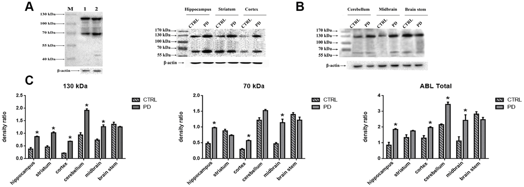
Figure 3.Detection of altered protein glycosylation in various mouse brain areas. (A) Western blot analysis of protein glycosylation in whole brain using ABL, which selectively labels Gal-(β-1,3)-GalNAc oligosaccharides: lane 1, control; lane 2, PD. (B) Western blot analysis of protein glycosylation in the indicated brain areas. (C) Densitometric analysis of Gal-(β-1,3)-GalNAc oligosaccharides levels normalized to the level of β-actin. Bars represented the mean±SD; * p<0.05 vs. control.