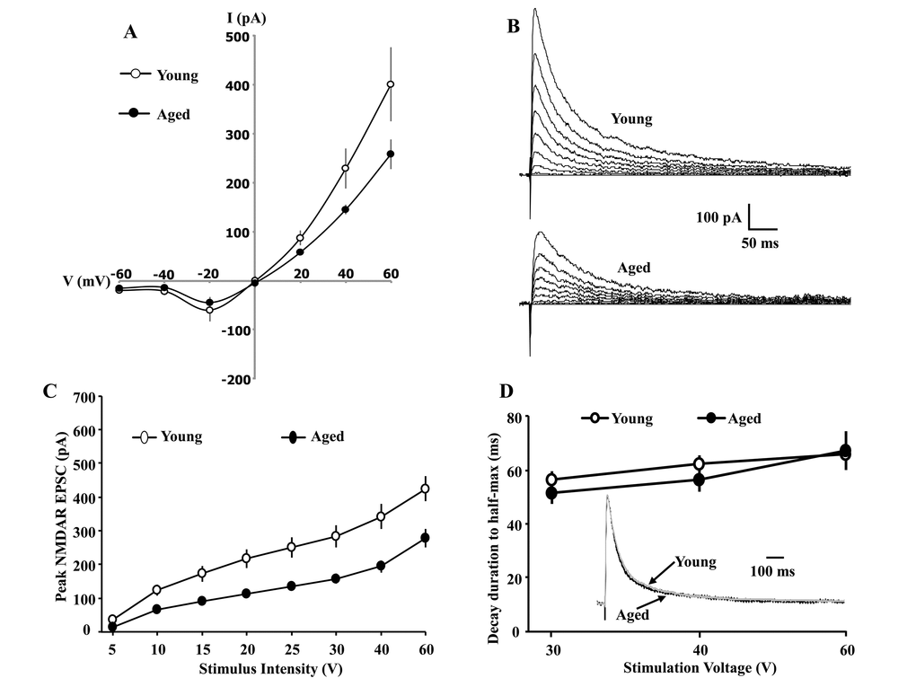
Figure 1.Whole-cell patch clamp recording from CA1 hippocampal pyramidal neurons of aged and young animals demonstrating the current-voltage relationship and synaptic decay duration. (A) The current-voltage relationship was recorded from CA1 pyramidal neurons from young (11/4 cells/animals) and aged (9/4 cells/animals) animals. When cells are clamped at positive voltages, the currents are outward and larger currents are observed for young animals. The reversal potential is near 0 mV for both age groups. When cells are clamped at negative voltages, currents are inward and reduced, consistent with Mg2+ blockade of the NMDAR channel. Examination of peak amplitude and time to half-decay of the NMDAR EPSC during aging. The cells were voltage clamped at +40 mV. (B) Representative traces evoked by the eight different stimulation intensities and recorded from young (top) and aged animals (bottom). (C) A decrease in the peak NMDAR EPSC was observed across the range of stimulation intensities for CA1 pyramidal cells recorded from aged animals (filled circle, n = 26/14 cells/animals), relative to cells from young animals (open circle, n = 20/9 cells/animals). (D) The mean (±SEM) time for the EPSC to decay to 50% of the peak for the three highest stimulation intensities. The inset shows the time course of the EPSC, evoked by 40 V stimulation, across all CA1 pyramidal cells recorded from young (gray trace, n = 20/9 cells/animals) and aged (dark trace, n = 26/14 cells/animals) animals. For each cell, the response amplitude evoked by 40 V stimulation was normalized to the peak of the response.