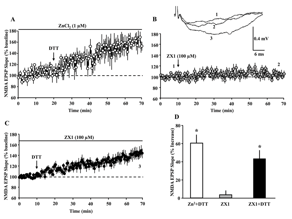
Figure 5.The DTT-induced potentiation of the NMDAR-synaptic response is not due to zinc chelation. The panels A-C illustrate the time course for the NMDAR-fEPSP slope; each point represents the mean (±SEM), normalized to the baseline (dashed line). (A) The arrow indicates the time of bath application of DTT (0.5 mM) in presence of ZnCl2 (1 µM). (B) The arrow indicates the time of bath application of ZX1 (100 µM). (C) The last ten min of NMDAR-fEPSP slope recording in presence of ZX1 was renormalized and DTT was added (arrow). (D) Bar graph represents the mean (+SEM) percent change in NMDAR-mediated fEPSP during the last 5 min of recording, in response to Zn2+ plus DTT (open bar, n = 3/3 slices/animals), ZX1 alone (gray bar, n = 11/6 slices/animals,) and ZX1 plus DTT (filled bar, n = 11/6 slices/animals). Asterisks indicate significant potentiation relative to baseline.