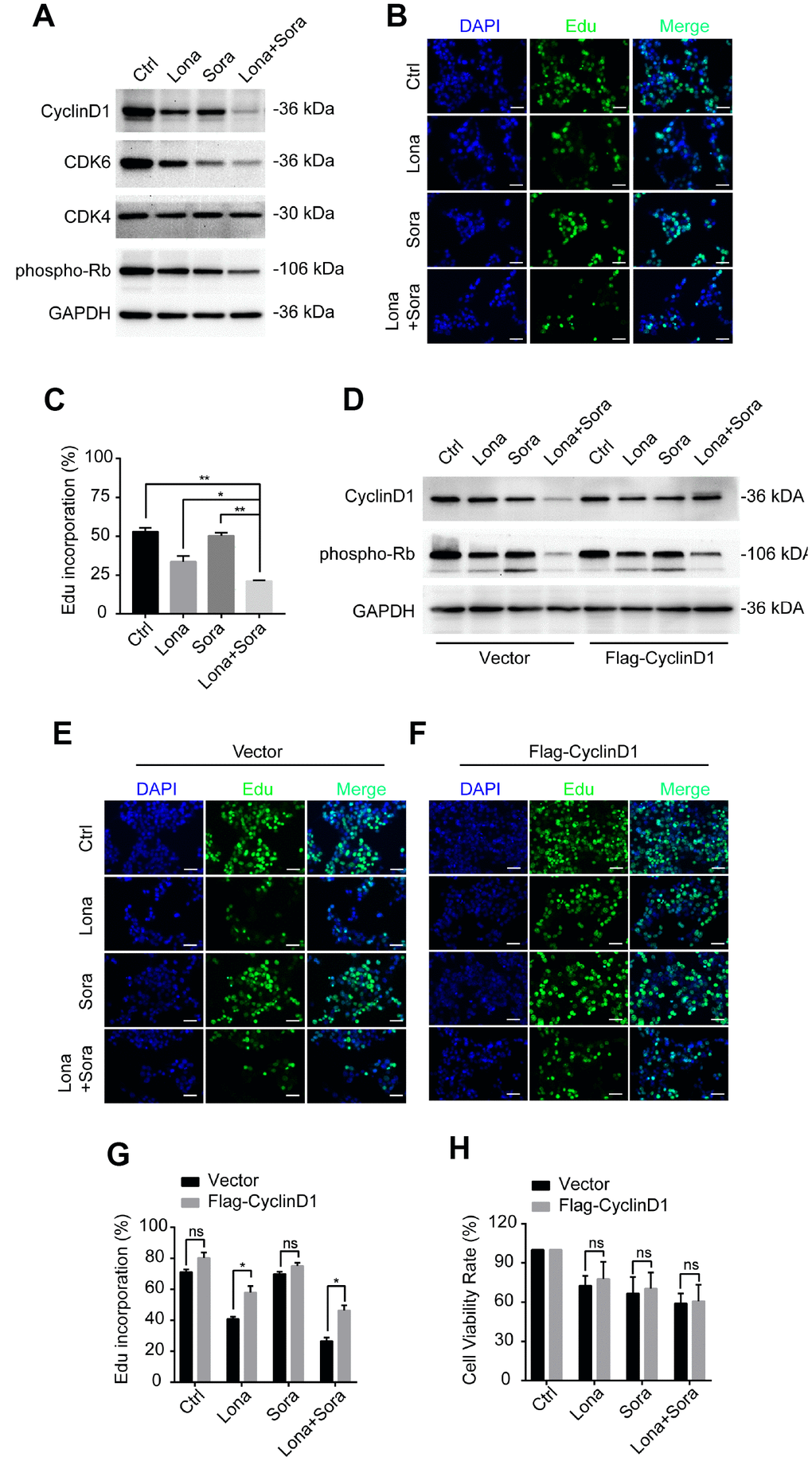
Figure 7.Degradation of cyclin D1 caused by lonafarnib and sorafenib co-treatment inhibits DNA synthesis. (A) HepG2 cells were treated with lonafarnib and/or sorafenib for 48 h. Protein expression levels were determined using the antibodies as indicated. (B) HepG2 cells were treated with lonafarnib and/or sorafenib for 48 h. DNA synthesis was analyzed by Edu staining. (C) Quantification of Edu positive straining in (B) was shown in the diagram.. *P < 0.05; **P < 0.01. (D) Protein expression levels of cyclin D1 and phospho-Rb in HepG2 cells transfected with vector control or Flag-Cyclin D1. (E and F) Representative image of Edu staining in HepG2 cells transfected with vector control or Flag-Cyclin D1. (G) Quantification of Edu positive straining in (E) and (F). ns, P > 0.05; *P < 0.05. (H) HepG2 cells were treated with lonafarnib and/or sorafenib for 48 h, and cell viability was evaluated by CCK-8 assay. ns, P > 0.05.