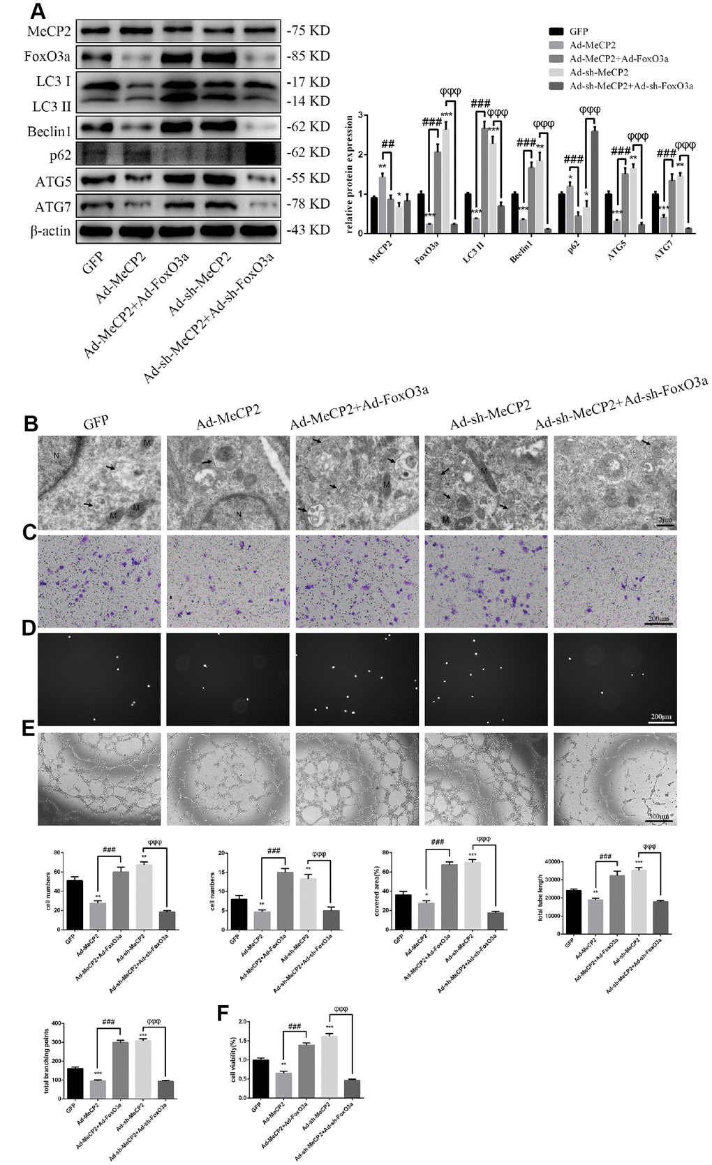
Figure 7.MeCP2 inhibits autophagy and EPC function through FoxO3a. (A) Protein levels of MeCP2, FoxO3a, LC3 II, Beclin1, p62, ATG5, and ATG7 were detected by western blotting after transfection with Ad-MeCP2 or Ad-sh-MeCP2 or co-transfection with Ad-FoxO3a or Ad-sh-FoxO3a for 48 h. (B) EPC ultrastructure was imaged by TEM. The white arrow indicates autophagosomes. N, nucleus; M, mitochondria. (C) Cell migration was evaluated by Transwell migration assays after transfection or co-transfection for 48 h. (D) Cell adhesion ability was evaluated by matrix adhesion assay after transfection or co-transfection for 48 h. (E) Angiogenic ability was evaluated by Matrigel assays after transfection or co-transfection for 48 h. (F) Cell viability was evaluated with CCK-8 after transfection or co-transfection for 48 h. *P < 0.05, **P < 0.01, and ***P < 0.001 vs. control. #P < 0.05, ##P < 0.01, and ###P < 0.001 vs. Ad-MeCP2 treatment. φP < 0.05, φφP < 0.01, and φφφP < 0.001 vs. Ad-sh-MeCP2 treatment.