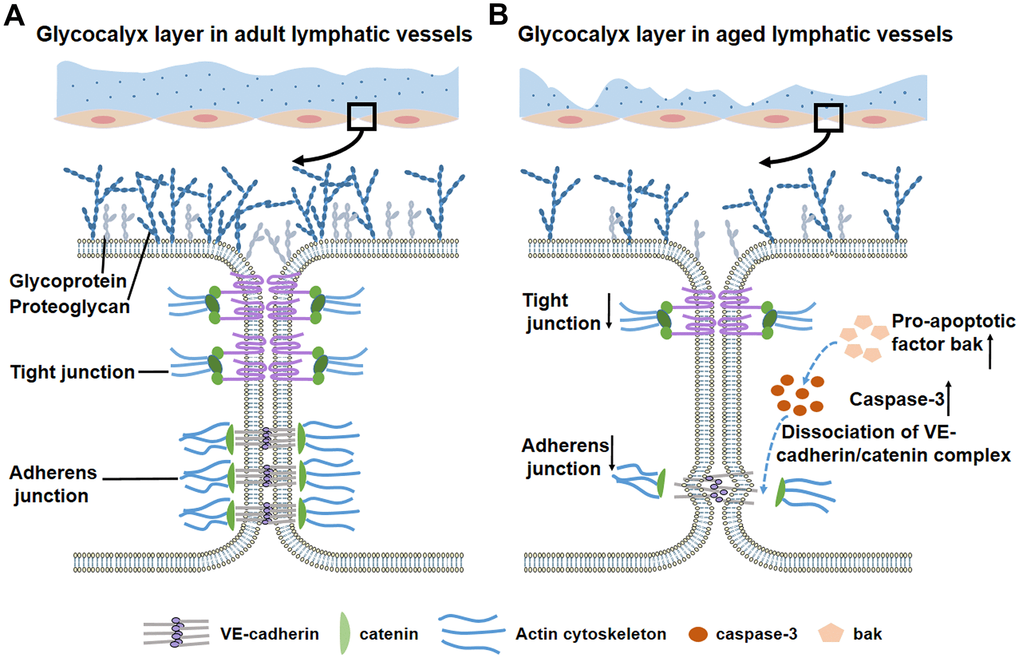
Figure 3.Glycocalyx layer and intercellular junctions of lymphatic vessels during the aging process. (A) In adult lymphatic vessels, the intact, continuous glycocalyx layer covers lymphatic endothelial cells. Detailed view in the box shows the normal glycocalyx layer and intercellular junctions. (B) Aged lymphatic vessels display thin, discontinuous glycocalyx layer. Detailed view in the box shows a significant loss of glycocalyx and adherens/tight junctions. Increased pro-apoptotic factor bak activates caspase-3 to disrupt the downstream protein β-catenin, which leads to decreased adherens junctions and impaired barrier function.