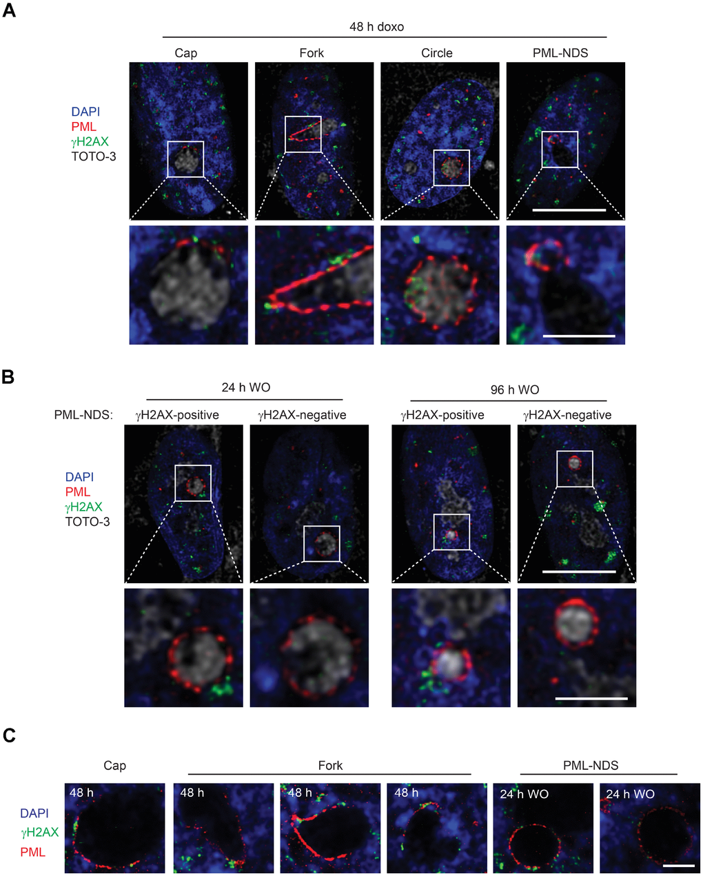
Figure 6.PNAs colocalize with persistent DNA damage foci. Co-association of DNA damage foci and PNAs detected by γH2AX (green) and PML (red) immunostaining, respectively, in RPE-1hTERT treated with 0.75 μM doxorubicin for 48 hours (A) and 24 and 96 hours after drug removal (B). Higher magnifications of confocal microscopic images of PML/γH2AX co-associations (insets) are shown in the lower rows. Bars, 10 μM for the whole cells and 3 μM for the insets. (C) High resolution STED microscopic images of PNAs/γH2AX co-associations in RPE-1hTERT treated with 0.75 μM doxorubicin for 48 hours and followed for PML-NDS appearance 24 hours after drug removal (WO). Nuclei are stained with DAPI (blue). Bars, 4 μM.