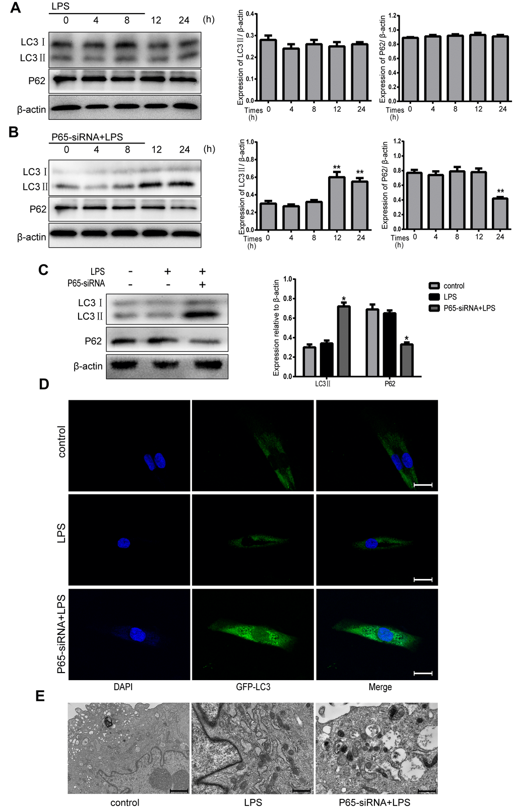
Figure 4.Inhibiting NF-κB promotes autophagy in LPS-induced NPCs. (A) After treatment with LPS for 4-24 h, the protein expression levels of LC3 II and P62 were measured by western blot. (B) After treatment with LPS plus p65-siRNA for 4-24 h, the protein expression levels of LC3 II and P62 were measured by western blot. (C) Western blot analysis for the protein expression levels of LC3 II and P62 after various treatment for 24 h. (D) NPCs were transfected with adenovirus containing GFP-LC3 and the formation and distribution of GFP-LC3 punctate were observed under confocal microscopy (Amplification×800). (E) Morphological observation of autophagy under transmission electron microscope (magnification ×30,000). Values are means ±SEM.*p<0.05 vs. control group, #p<0.05 vs. LPS group, **p<0.05 vs. only p65-siRNA group.