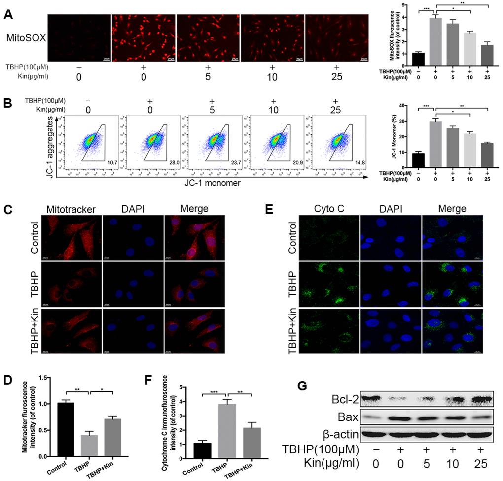
Figure 2.Kin attenuates TBHP-induced mitochondrial dysfunction in NPCs. (A) The mitochondria-derived ROS in NPCs, treated with different concentrations of Kin (0, 5, 10 or 25 μg/ml) for 2 h before receiving TBHP (100 μM) for 24 h, was detected by MitoSOX staining, and the red fluorescence intensity was quantified; scale bar: 20 μm. (B) The mitochondrial membrane potential in NPCs, treated with different concentrations of Kin (0, 5, 10 or 25 μg/ml) for 2 h before receiving TBHP (100 μM) for 24 h, were analyzed by flow cytometry using JC-1 staining. (C, D) The NPCs were treated with TBHP (100 μM) alone for 24 h, or Kin (25 μg/ml) for 2 h before receiving TBHP (100 μM) for 24 h. The mitochondrial membrane potential was measured by Mitotracker staining and the fluorescence intensity was quantified; scale bar: 10 μm. (E, F) The immunofluorescence staining of Cyto C in NPCs; scale bar: 10 μm. (G) The western blotting of Bcl-2 and Bax in the NPCs treated with different concentrations of Kin (0, 5, 10 or 25 μg/ml) for 2 h before receiving TBHP (100 μM) for 24 h. All data are expressed as mean ± SD of at least three independent experiments.