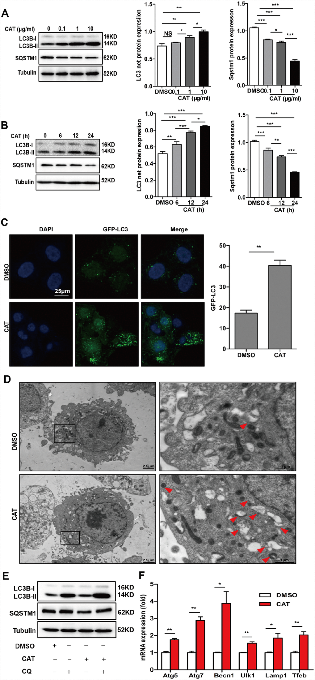
Figure 3.CAT induced autophagy in hepatocytes. (A) Dose-dependent induction of autophagy by CAT. Representative immunoblot analysis of LC3-II and SQSTM1 expression in lysates from HepG2 cells that were treated with CAT or control solvent (dimethyl sulfoxide, DMSO) at the indicated concentrations for 24 h. (B) Time-dependent induction of autophagy by CAT. Immunoblot detection of LC3-II and SQSTM1 expression in HepG2 cells treated with CAT (10 μg/mL) or DMSO for the indicated time. (C) HepG2 cells were transduced with an GFP-LC3 plasmid for 48 h and then treated with CAT (10 μg/mL) for 24h. Cells were observed by fluorescence microscopy to evaluate the number expressing GFP-LC3. (D) Representative electron microscopic pictures of HepG2 cells treated with CAT for 24 h. Arrows indicate autophagosomes. (E) Immunoblot analysis of LC3-II and SQSTM1 expression in HepG2 cells treated with 10 μg/mL CAT for 24 h in the absence or presence of 50 mM chloroquine (CQ) for the last 2 h. (F) qPCR analysis of autophagy-related and lysosomal genes in CAT-treated HepG2 cells. Means ± SD were calculated from three independent experiments. *P < 0.05, **P < 0.01.