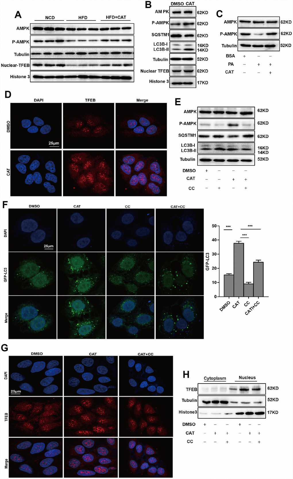
Figure 4.CAT induced autophagy via the AMPK-TFEB pathway. (A) Immunoblot detection of TFEB, AMPK, and AMPK phosphorylation levels in livers of mice. (B) Immunoblot detection of LC3-II, SQSTM1, TFEB, AMPK, and AMPK phosphorylation levels in HepG2 cells treated with CAT (10 μg/mL) for 24 h. (C) Immunoblot analysis of AMPK and p-AMPK levels in HepG2 cells treated with 0.3 mM palmitate (PA) and 10 μg/mL CAT for 24 h. (D) Fluorescence microscopy images of nuclear TFEB in HepG2 cells treated with 10 μg/mL CAT for 24 h. Scale bars: 25 μm. (E) Immunoblots of LC3-II, SQSTM1, AMPK, and AMPK phosphorylation in HepG2 cells treated with CAT (10 μg/mL) and Compound C (CC, 10 μM) for 24 h. (F) Numbers of GFP-LC3 in HepG2 cells expressing GFP-LC3 after treatment with or without CAT (10 μg/mL) and CC (10 μM) for 24 h were evaluated using fluorescence microscopy. Scale bars: 25 μm. (G, H) Fluorescence microscopy images and Immunoblot analysis of nuclear TFEB in HepG2 cells treated with 10 μg/mL CAT and Compound C (CC, 10 μM) for 24 h. Scale bars: 25 μm. ***P < 0.001.