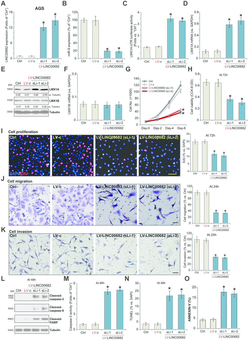
Figure 1.Ectopic overexpression of LINC00682 induces miR-9 downregulation but LMX1A upregulation, inhibiting AGS cell survival, proliferation, migration and invasion. AGS cells were infected with LINC00682-expressing lentivirus (“LV-LINC00682”), following puromycin selection two stable lines (“sLi-1/-2”) were obtained; Control cells were infected with the lentivirus with empty vector (“LV-c”); In those cells expression of LINC00682 (A), miR-9 (B), LMX1A mRNA (D), LMX1B mRNA (F) was tested by qPCR; The relative LMX1A3’-UTR luciferase activity was tested (C); Expression of the listed proteins in total cell lysates was tested by Western blotting (E); Cells were further cultured for the indicated time periods, cell survival, proliferation, migration and invasion in vitro were tested by the appropriate assays (G–K); Cell apoptosis was tested by Western blotting assay of apoptosis proteins (L), caspase-3 activity assay (M), nuclear TUNEL staining assay (N) and Annexin V FACS staining (O). The exact same number of viable cells of different genetic treatments were plated initially (“0h”/“Day-0”) for the functional assays (Same for all following Figures). Five repeated views in each condition were included to calculate the average number of migrated/invasive cells (Same for all Figures). Listed proteins were quantified and normalized to the loading control (E). “MW” stands for molecular weight (Same for all Figures). “Ctrl” stands for the parental control cells (Same for all Figures). For each assay, n=5 (five dishes or wells). *P <0.05 vs. “LV-c” cells. Experiments in this figure were repeated four times, and similar results were obtained. Bar=100 μm (I, J and K).