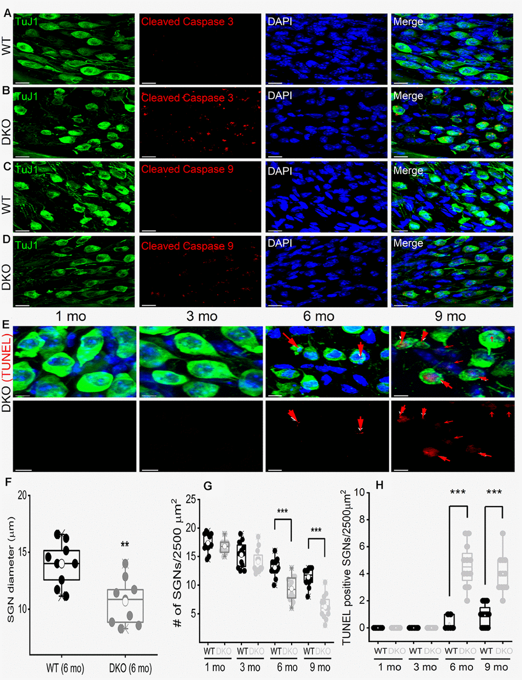
Figure 11.Age-related degeneration of SGN through apoptosis signal. (A–D) Immunofluorescence detection of cleaved caspase 3 (red; A–B) and caspase 9 (red; C, D) in WT and DKO 5-mo old cochlear sections. SGNs were labeled with neuronal marker TuJ1 (green). The nuclei were stained with DAPI (blue). Scale bar: 20 μm. Increased active caspase 3/9 was seen in DKO cochlear sections. Scale bar = 10 μm. (E) Shown are cochlear sections of in DKO cochleae at difference ages (1-9 mos) assessed for with TUNEL assay (TUNEL-positive in red). Scale bar: 5 μm. (F) Summary histogram showing a significant reduction in SGN size (diameter) between WT and DKO mice at 6-mo old (data from 9 mice each). (G) Age-dependent (1-9 mo) reduction in SGN densities (data from 8-9 mice each). Comparison between WT and DKO mice. (H) Summary data from WT and DKO cochleae at ages indicated, showing increased TUNEL-positive SGNs in 6-9–mo old (data from 8-9 mice each). Significant differences between genotypes were determined using unpaired two-tailed t-test (* p < 0.05, ** < 0.01, *** < 0.001).