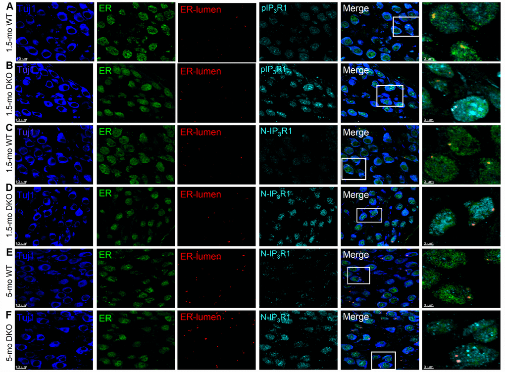
Figure 12.Detection of IP3R1, pIP3R1, and its proteolytic fragments in SGNs from WT and DKO cochlea. (A–F) Immunofluorescence detection of the endoplasmic reticulum (ER)-lumen domain of IP3R1(red), phosphorylation site (pIP3R1) (cyan; A–C), N-terminal domain (N-IP3R1) (cyan; C–F). We examined basal cochlear sections at 1.5-mo and 5-mo WT and DKO mice. SGNs were labeled with neuronal marker TuJ1 (blue). The ER was stained with ER-marker (blue). Merged images are shown together with digitally magnified (~3X) images, shown in the last panel. Scale bar 3 μm. For pIP3R1 at 1.5 mos, the percent of SGNs with positive reactivity for WT was 12 ± 3, and DKO was 54 ± 9; (p < 0.0001, data from 7 mice obtained from 25 section/mice). N-IP3R1 at 1.5- and 5-mos, percent cell reactivity for WT = 6 ± 3; DKO = 37 ± 12, and WT = 9 ± 4; DKO = 61 ± 12, respectively (p < 0.0001, data from 6 mice obtained from 25 section/mice). Scale bar = 10 μm.