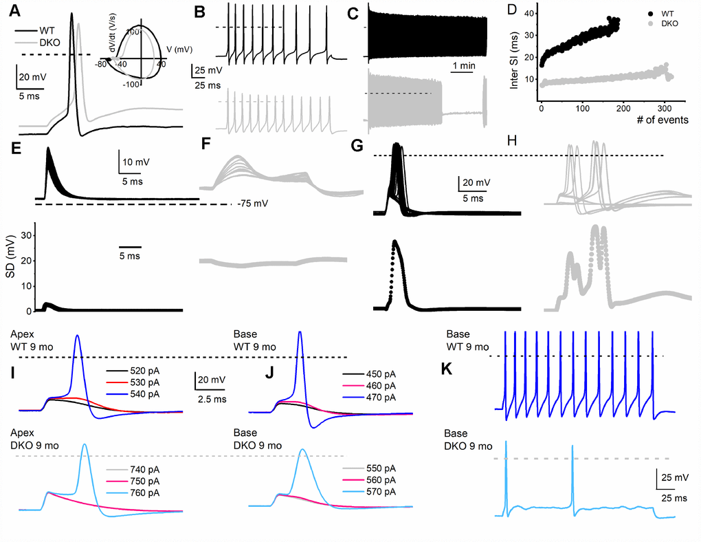
Figure 4.Membrane properties of SGNs from WT and DKO mice. To minimize experimental variabilities, SGN recordings were performed using mice, with recorded ABR. Current-clamp recordings were performed on SGNs isolated from the basal and apical one-third of the cochlea from 1-, 3-, 4-5- (not shown), 6- and 9-mo old WT and DKO mice. (A) Action potentials (AP) evoked from 1-mo-old SGNs isolated from cochlear apical turn from WT (black trace) and DKO (grey trace) mice. Both the resting membrane potential (RMP) and AP amplitude were significantly altered in WT versus DKO SGNs. RMP of apical SGNs; WT, -65 ± 3 mV, n = 31; DKO, -58 ± 2 mV, n = 27; p < 0.001: RMP of basal SGNs; WT, -57 ± 2 mV, n = 25; DKO, -53 ± 4 mV, n = 31; p < 0.001). AP amplitudes were; apical SGN; WT, 80.0 ± 2.7 mV, n = 15; DKO, 72.1 ± 4.8 mV, n = 15; p < 0.001: basal SGN; WT, 83.2 ± 3.4 mV, n = 17; DKO, 72.0 ± 5.1 mV, n = 19; p < 0.001). Dotted black and gray lines indicate 0 mV. (B, C) Examples of slow adapting SGNs isolated from a 1-mo-old basal cochlear turn. For the example shown in (B) typical spike frequency (in Hz) for WT SGNs = 45 ± 7 (n = 17) and DKO = 67 ± 11 (n = 21); p < 0.0001. (D) The dairy plot of the inter-spike interval of slow adapting SGNs from WT and DKO. (E, F) 50 consecutive subthreshold depolarization (0.075 nA current injection) of WT SGNs (E, black traces), and DKO (F, grey traces), demonstrating the extent of membrane jitters. Plotted below is the corresponding standard deviation. (G, H) 30 consecutive suprathreshold depolarization (0.2 nA current injection; interstimulus interval, 2s) of WT SGNs (G, black traces), and DKO (H, grey traces), demonstrating the extent of membrane jitters in evoked APs. Plotted below is the corresponding standard deviation. DKO membrane voltage is pre-disposed to increased membrane jitters. (I, J) APs evoked in SGNs from 9-mo old WT (upper panel) cochlea were generally slower to initiate and larger in amplitude compared to those evoked in SGNs from DKO (lower panel) cochlea. The magnitudes of the injected current are indicated. (K) Across SGNs, the excitability of DKO SGNs has plummeted by several-fold. For the example shown, the spike frequency was reduced by ~7-fold.