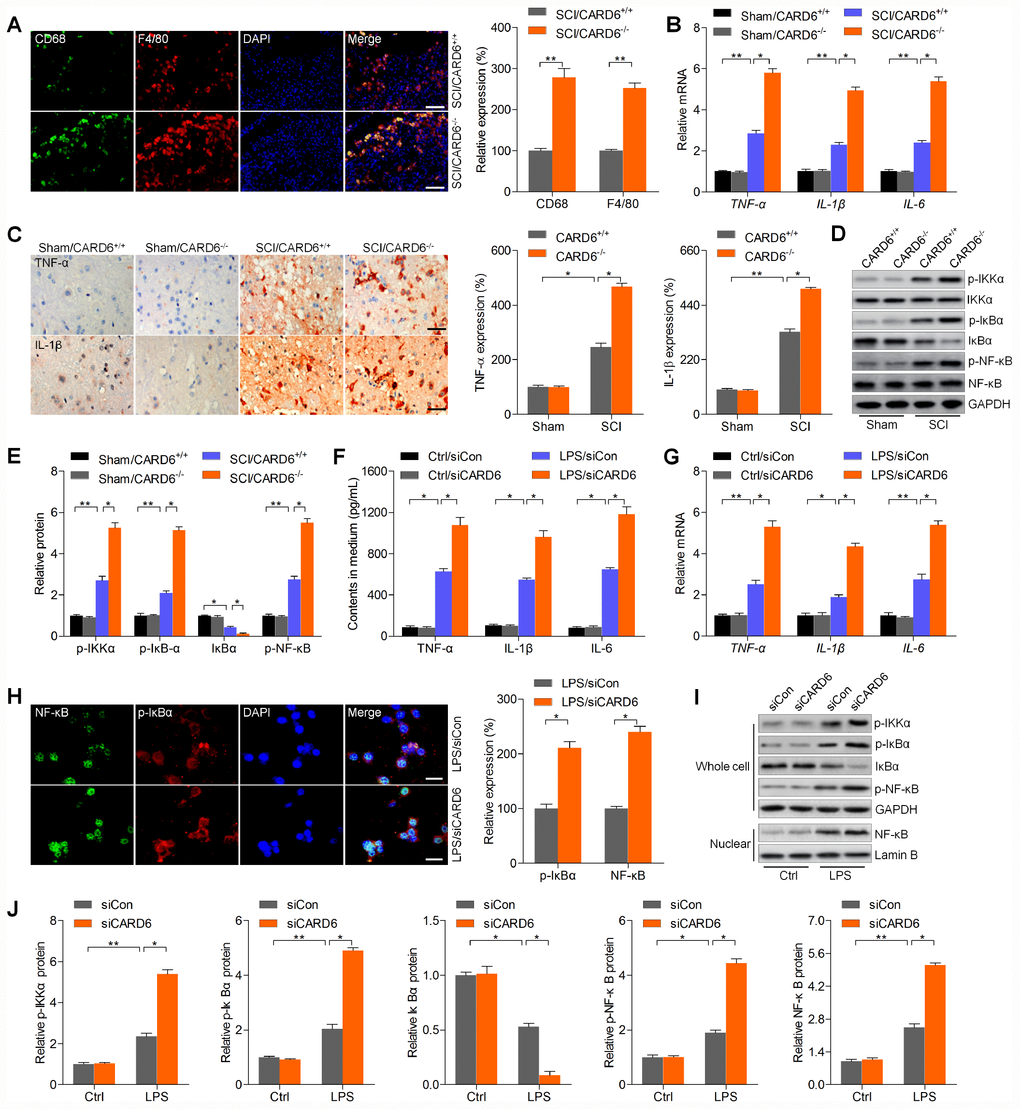
Figure 4.CARD6 knockout accelerates inflammatory response in mice after SCI. (A) Representative images of CD68/F4/80 double staining by IF in dorsal horn of mice. The relative expression of CD68 and F4/80 was quantified. Scale bar: 100 μm. (B) RT-qPCR analysis of TNF-α, IL-1β and IL-6 mRNA levels in the lumbar spinal cord segments. (C) Representative images of TNF-α and IL-1β and by IHC staining in dorsal horn of mice. The relative expression of TNF-α and IL-1β was quantified. Scale bar: 100 μm. (D, E) Western blot analysis of p-IKKα, p-IκBα, IκBα and p-NF-κB protein expression levels in the lumbar spinal cord segments. (F–J) BV2 cells were transfected with siCARD6 or siCon for 24 h, followed by LPS exposure for another 24 h. Then, all cells were collected for further studies. (F) TNF-α, IL-1β and IL-6 contents in medium were assessed by ELISA. (G) TNF-α, IL-1β and IL-6 mRNA levels in cells were measured using RT-qPCR analysis. (H) Representative images of p-IκBα and NF-κB double staining by IF in cells. The quantification of p-IκBα and NF-κB expression levelv was exhibited. Scale bar: 50 μm. (I, J) Protein expression levels of p-IKKα, p-IκBα, IκBα and p-NF-κB in whole cells, and NF-κB in nuclear were determined by western blot analysis. Data represented means ± SEM (n=8 each group for in vivo studies; n=6 each group for in vitro studies). *p < 0.05 and **p < 0.01.