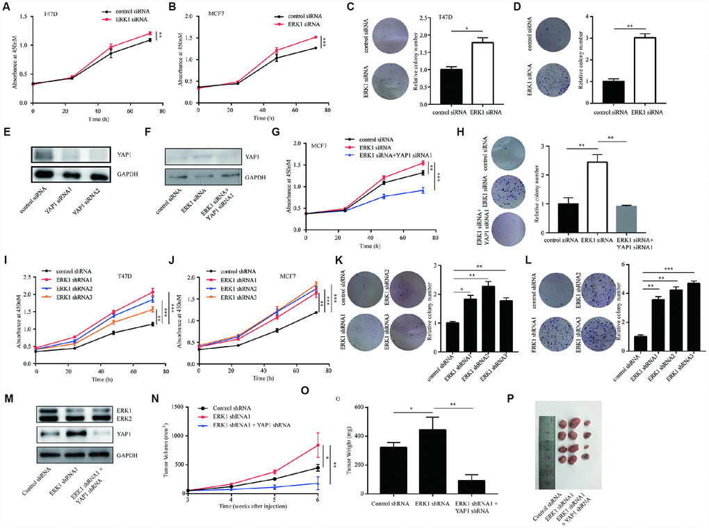
Figure 5.ERK1 promoted cell proliferation of breast cancer cells in vitro and in vivo. (A) In T47D cells, silencing of ERK1 increased cell proliferation ability. (B) In MCF7 cells, silencing of ERK1 increased cell proliferation ability. (C) In T47D cells, silencing of ERK1 increased colony forming ability. (D) In MCF7 cells, silencing of ERK1 increased colony forming ability. (E) Transfection of YAP1 siRNA decreased YAP1 protein expression in MCF7 cells. (F) Silencing of YAP1 reversed ERK1 silencing induced elevation of YAP1 protein expression in MCF7 cells. (G) Silencing of YAP1 reversed ERK1 silencing induced elevation of cell proliferation ability in MCF7 cells. (H) Silencing of YAP1 reversed ERK1 silencing induced elevation of colony forming ability in MCF7 cells. (I) The cell proliferation of T47D cells with stable knockdown of ERK1 was increased in comparison with T47D cells infected with control shRNA. (J) The cell proliferation of MCF7 cells with stable knockdown of ERK1 was increased in comparison with MCF7 cells infected with control shRNA. (K) The colony forming ability of T47D cells was decreased after lentivirus mediated ERK1 knockdown. (L) The colony forming ability of MCF7 cells was decreased after lentivirus mediated ERK1 knockdown. (M) Western blotting showed that lentivirus mediated knockdown of ERK1 decreased ERK1 protein expression and elevated YAP1 protein expression, while knockdown of both ERK1 and YAP1 decreased ERK1 and YAP1 protein expression in MCF7 cells. (N) Xenografted tumor growth curve indicated that ERK1 knockdown increased tumor volume, while YAP1 knockdown decreased tumor volume in vivo. (O) ERK1 knockdown increased tumor weight, while YAP1 knockdown decreased tumor weight in vivo. (P) Representative xenografted tumors from nude mouse models. *, p<0.05; **, p<0.01; ***, p<0.001.