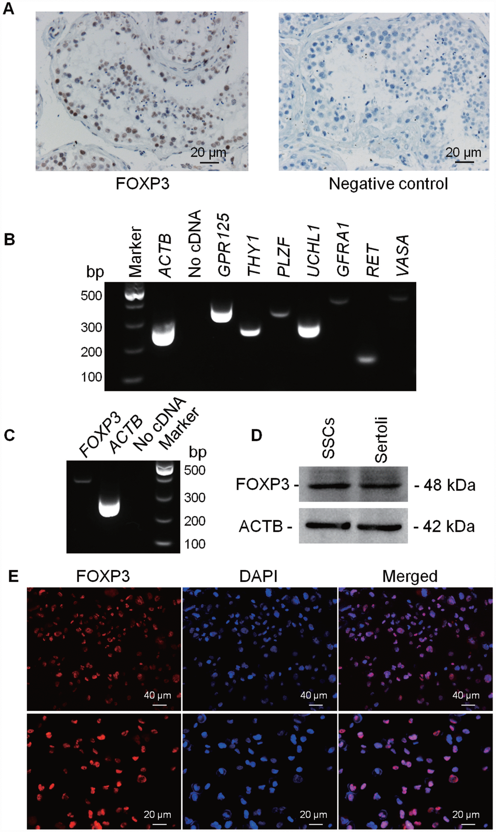
Figure 4.Expression and location of FOXP3 proteins in human testes and human SSC line. Immuno-histochemistry revealed cellular localization of FOXP3 in human testes (left panel). Replacement of anti-FOXP3 with PBS was used as a negative control (right panel). Scale bars = 20 μm. (B) RT-PCR showed the transcripts of GPR125, THY1, PLZF, UCHL1, GFRA1, RET and VASA in human SSC line. Samples without cDNA (No cDNA) but PCR with gene primers were used as negative controls. ACTB served as loading controls of total RNA. (C) RT-PCR showed the mRNA level of FOXP3 in human SSC line. Samples without cDNA (No cDNA) but PCR with gene primers were used as negative controls, and ACTB served as a loading control of total RNA. (D) Western blots revealed the expression of FOXP3 protein in human SSC line. ACTB served as the control of the loading proteins. Human Sertoli cells were utilized as a positive control. (E) Immunocytochemistry revealed cellular localization of FOXP3 in human SSC line. Fluorescent signals of FOXP3 (red) and DAPI (blue) were imaged individually and merged under fluorescence microscope. Scale bars = 40 μm and 20 μm, respectively. All experiments were repeated for at least three times.