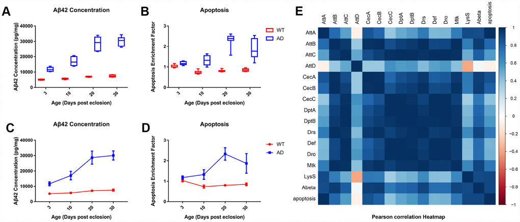
Figure 4.Quantitative determination of Aβ42 and apoptosis levels in brain tissue of control and AD model flies. Aβ42 concentration and apoptotic DNA fragmentation representing the extent of apoptosis was examined with ELISA. Compared to the control group (left), which had low levels of Aβ42 concentration (A, left) and cell apoptosis (B, left), the AD group had an increased concentration of Aβ (A, right) and apoptotic DNA fragments (B, right). The trends of Aβ42 production (C) and neuronal apoptosis (D) in the WT (round dots) and AD (square dots) groups are displayed in the lower figures. Subsequent correlation analysis further indicated a potential relationship between AMP expression, Aβ42 production, and neuronal apoptosis.