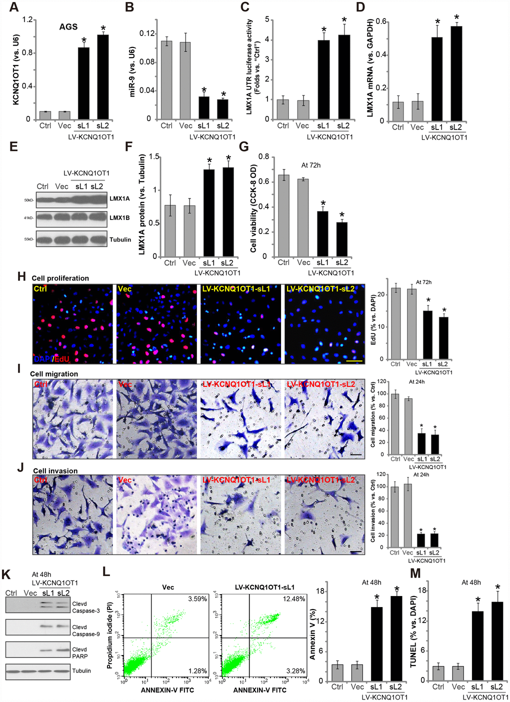
Figure 2.Forced overexpression of LncRNAKCNQ1OT1 induces miR-9 depletion, LMX1A upregulation and AGS cell inhibition. AGS cells were infected with KCNQ1OT1-expressing lentivirus (“LV-KCNQ1OT1”) or scramble control vector lentivirus (“Vec”) for 24h, following puromycin selection two stable lines (“sL1/sL-2”) with LV-KCNQ1OT1 established, expression of KCNQ1OT1 (A), miR-9 (B) and LMX1A mRNA (D) were tested by qPCR assays; The LMX1A 3’-UTR luciferase activity was shown (C); Expression of the listed proteins in total cell lysates were tested by Western blotting assay (E, results quantified in F); Cells were further cultured for the indicated time periods, cell survival and proliferation were tested by CCK-8 assay (G) and EdU staining (H), respectively; Cell migration and invasion were tested by “Transwell” (I) and “Matrigel Transwell” assay (J), respectively; Cell apoptosis was tested by Western blotting (testing apoptosis-associated proteins, K), Annexin V FACS (L) and TUNEL staining assay (M). The exact same number of viable cells of different genetic treatment were plated initially (at 0h) for the functional assays (Same for all following Figures). “Ctrl” stands for the parental control cells (Same for all Figures). For each assay, n=5. *P <0.05 vs. “Vec” cells. Experiments in this figure were repeated five times, and similar results were obtained. Bar=100 μm (H–J).