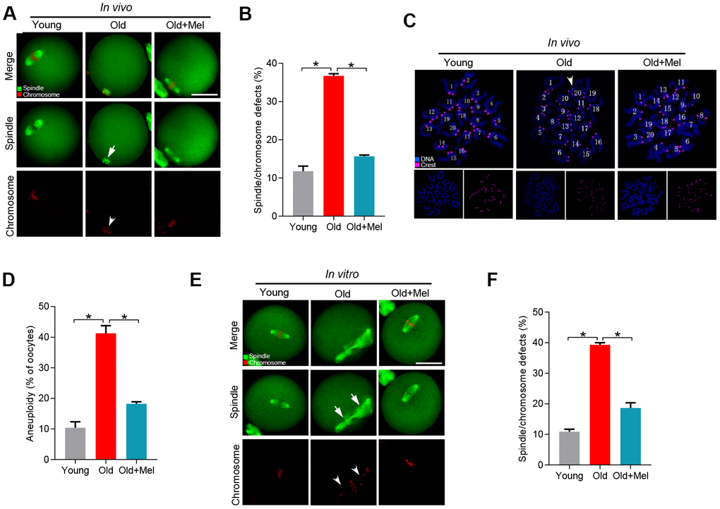
Figure 1.Effects of melatonin administration on meiotic apparatus in aged oocyte. (A) MII oocytes from young (n=90), old (n=88) and old mice treated with melatonin (old+Mel, n=80) were stained with α-tubulin to visualize spindle (green) and counterstained with propidium iodide to visualize chromosomes (red). Representative confocal sections are shown. Arrows indicate the spindle defects and arrowheads point to the misaligned chromosomes. (B) Quantification of young, old and old+Mel oocytes with abnormal spindle and chromosomes. (C) Chromosome spreading of young, old and old+Mel MII oocytes with aneuploidy. Chromosomes were stained with Hoechst 33342 (blue), and kinetochores were labeled with CREST (purple). (D) Histogram showing the incidence of aneuploidy in young (n=82), old (n=57) and old+Mel (n=53) oocytes. (E) Young and old oocytes in vitro cultured with or without melatonin were processed to evaluate meiotic apparatus. Young, old, and old+Mel oocytes were stained with α-tubulin to visualize spindle (green) and counterstained with propidium iodide to visualize chromosomes (red). Representative confocal sections are shown. Arrow indicates the disorganized spindle and arrowhead indicates the misaligned chromosomes. (F) Quantification of young (n=131), old (n=78) and old+Mel (n=95) oocytes with abnormal spindle/chromosomes. Data are expressed as mean percentage ± SD from three independent experiments. *P<0.05 vs. controls. Scale bar: 50 μm.