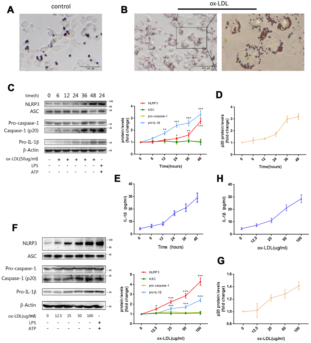
Figure 1.Ox-LDLs activate NLRP3 inflammasomes in a time- and dose-dependent manner. (A and B) Representative Oil Red O staining images of Mφ treated with (B) or without (A) ox-LDLs. The right image in (B), 4× enlargement of the outlined area at the left. Scale bars, 100 um. (C) The immunoblot analysis of lysates of Mφ treated with ox-LDLs (50 ug/ml) for a series of time intervals. (C and D) The densitometric analysis of the NLRP3, ASC, pro-caspase-1, pro-IL-1β (C) and p20 (D) signal vs. time, which was normalized to β-actin. (E) The ELISA of IL-1β in supernatants obtained from (C). (F) The immunoblot analysis of lysates of Mφ treated with various doses of ox-LDLs for 24 hours. (F and G) The densitometric analysis of the NLRP3, ASC, pro-caspase-1, pro-IL-1β (F) and p20 (G) signal vs. ox-LDL concentrations, which were normalized to β-actin. (H) The ELISA of IL-1β in supernatants obtained from (F). The data are presented as mean ± SD (n=3); * denotes statistical significance by one-way analysis of variance (ANOVA) with post hoc Dunnett’s multiple comparisons test when compared to 0 hour or 0 ug/ml ox-LDLs. *P<0.05, **P<0.01, ***P<0.001.